Application
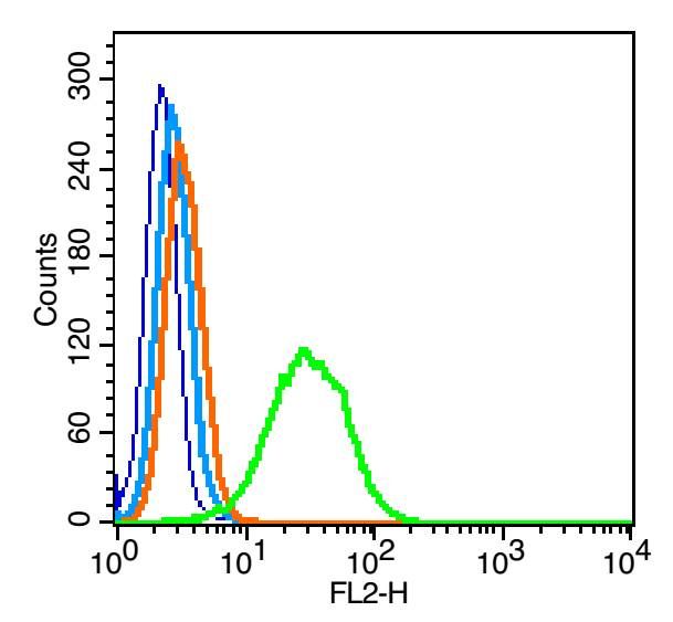
Fig1: Blank control: RSC96(blue), the cells were fixed with 2% paraformaldehyde (10 min) and then permeabilized with ice-cold 90% methanol for 30 min on ice. Isotype Control Antibody: Rabbit IgG(orange) ; Secondary Antibody: Goat anti-rabbit IgG-PE(white blue), Dilution: 1:200 in 1 X PBS containing 0.5% BSA ; Primary Antibody Dilution: 0.2μg in 100 μL1X PBS containing 0.5% BSA(green).Application
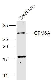
Fig2: Sample: Cerebrum (Mouse) Lysate at 40 ug Primary: Anti-GPM6A (ER1909-86) at 1/1000 dilution Secondary: IRDye800CW Goat Anti-Rabbit IgG at 1/20000 dilution Predicted band size: 31 kD Observed band size: 31 kD| Product Name | GPM6A |
|---|---|
| Antibody Type | Primary Antibodies |
| Antigen Alias | GPM6A GPM6A_HUMAN M6a M6 a M6-a Neuronal membrane glycoprotein M6-a. |
| Product description | Involved in neuronal differentiation, including differentiation and migration of neuronal stem cells. Plays a role in neuronal plasticity and is involved in neurite and filopodia outgrowth, filopodia motility and probably synapse formation. GPM6A-induced filopodia formation involves mitogen-activated protein kinase (MAPK) and Src signaling pathways. May be involved in neuronal NGF-dependent Ca(2+) influx. May be involved in regulation of endocytosis and intracellular trafficking of G-protein-coupled receptors (GPCRs); enhances internalization and recycling of mu-type opioid receptor. |
| Immunogen | KLH conjugated synthetic peptide derived from human GPM6A 151-250/278 |
| Clonality | Polyclonal |
|---|---|
| Isotype | IgG |
| Host Species | Rabbit |
| Tested Applications | WBFC |
| WB:1:500-2000: FC:1μg/Test: |
|
| Species Reactivity | HumanMouseRatDogRabbit |
| Concentration | 1mg/ml |
| Alternative Names | GPM6A GPM6A_HUMAN M6a M6 a M6-a Neuronal membrane glycoprotein M6-a. |
|---|---|
| Molecular Weight(MW) | 31 KDa |
| Cellular Localization | Cell membrane; Multi-pass membrane protein. Cell projection, axon. Cell projection, dendritic spine. Cell projection, filopodium. Note=Localizes to cholesterol-rich lipid rafts of the plasma membrane of hippocampal neurons. Localized to plasma membrane of cell bodies and neurites of hippocampal neurons. Localized in membrane protrusions (filopodia and spines) of primary hippocampal neurons. Localized to the growth cone edge membrane of elongating axons. |
| SwissProt ID | P51674 |
|---|
Application
Fig1: Blank control: RSC96(blue), the cells were fixed with 2% paraformaldehyde (10 min) and then permeabilized with ice-cold 90% methanol for 30 min on ice. Isotype Control Antibody: Rabbit IgG(orange) ; Secondary Antibody: Goat anti-rabbit IgG-PE(white blue), Dilution: 1:200 in 1 X PBS containing 0.5% BSA ; Primary Antibody Dilution: 0.2μg in 100 μL1X PBS containing 0.5% BSA(green).
Application
Fig2: Sample: Cerebrum (Mouse) Lysate at 40 ug Primary: Anti-GPM6A (ER1909-86) at 1/1000 dilution Secondary: IRDye800CW Goat Anti-Rabbit IgG at 1/20000 dilution Predicted band size: 31 kD Observed band size: 31 kD| Application Notes | WB:1:500-2000: FC:1μg/Test: |
|---|
| Form | Liquid |
|---|---|
| Storage Instructions | Store at -20℃ for one year. Avoid repeated freeze/thaw cycles. The lyophilized antibody is stable at room temperature for at least one month and for greater than a year when kept at -20℃. When reconstituted in sterile pH 7.4 0.01M PBS or diluent of antibody the antibody is stable for at least two weeks at 2-4℃. |
| Storage Buffer | 0.01M TBS(pH7.4) with 1% BSA, 0.03% Proclin300 and 50% Glycerol. |


