Application
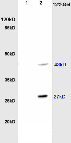
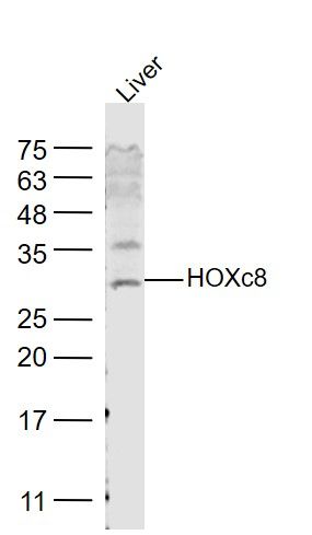
Fig1: Sample: Brain(Rat) lysate at 30ug; Liver(Rat) lysate at 30ug; Primary: Anti-HOXc8 (ER1910-77) at 1:200; Secondary: HRP conjugated Goat-Anti-Rabbit IgG(bse-0295G) at 1: 3000; Predicted band size : 27kD Observed band size : 27kD, 43kDApplication
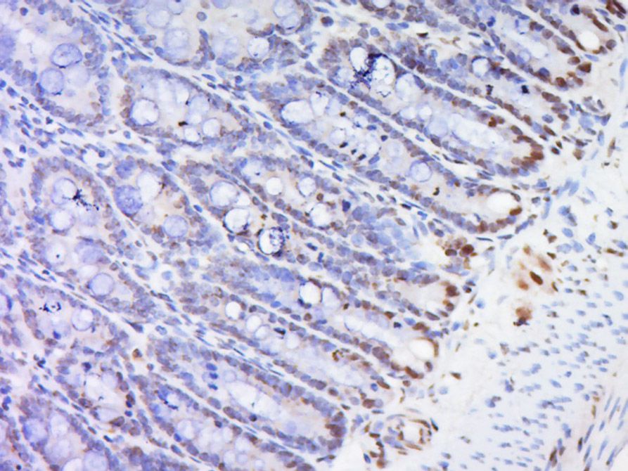
Fig2: Paraformaldehyde-fixed, paraffin embedded Rat small intestine; Antigen retrieval by boiling in sodium citrate buffer (pH6.0) for 15min; Block endogenous peroxidase by 3% hydrogen peroxide for 20 minutes; Blocking buffer normal goat serum at 37℃ for 30min; Antibody incubation with (Hox-3.1) Polyclonal Antibody, Unconjugated (ER1910-77) at 1:500 overnight at 4℃, followed by a conjugated secondary (sp-0023) for 20 minutes and DAB staining.Application
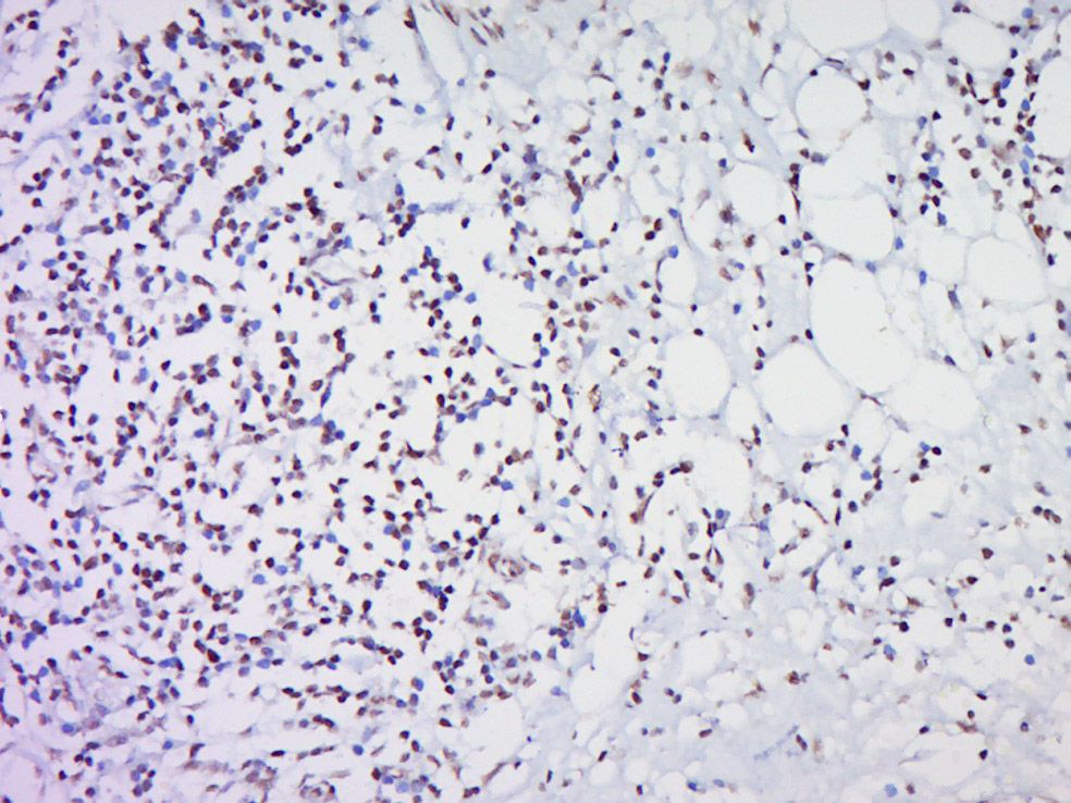
Fig3: Paraformaldehyde-fixed, paraffin embedded (Human cervical cancer); Antigen retrieval by boiling in sodium citrate buffer (pH6.0) for 15min; Block endogenous peroxidase by 3% hydrogen peroxide for 20 minutes; Blocking buffer (normal goat serum) at 37℃ for 30min; Antibody incubation with (Hox-3.1) Polyclonal Antibody, Unconjugated (ER1910-77) at 1:500 overnight at 4℃, followed by a conjugated secondary (sp-0023) for 20 minutes and DAB staining.Application
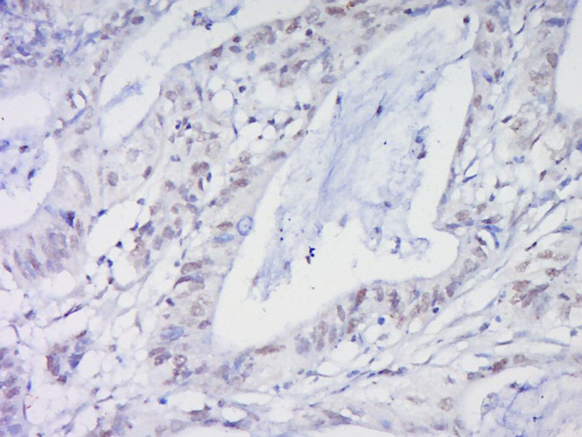
Fig4: Paraformaldehyde-fixed, paraffin embedded (Human cervical cancer); Antigen retrieval by boiling in sodium citrate buffer (pH6.0) for 15min; Block endogenous peroxidase by 3% hydrogen peroxide for 20 minutes; Blocking buffer (normal goat serum) at 37℃ for 30min; Antibody incubation with (Histone H3) Polyclonal Antibody, Unconjugated (ER1910-77) at 1:500 overnight at 4℃, followed by a conjugated secondary (sp-0023) for 20 minutes and DAB staining.Application
Fig5: Sample: Liver(Rat) Lysate at 40 ug Primary: Anti-HOXc8 (ER1910-77) at 1/500 dilution Secondary: IRDye800CW Goat Anti-Rabbit IgG at 1/20000 dilution Predicted band size: 27 kD Observed band size: 27 kD| Product Name | HOXc8 |
|---|---|
| Antibody Type | Primary Antibodies |
| Antigen Alias | Hox-3.1 Homeo box 3A Homeo box 8 Homeobox 3A Homeobox C8 Homeobox protein Hox C8 Homeobox protein Hox-C8 Homeobox protein HoxC8 Homeobox protein R4 Homolog of mouse Hox 3.1 HOX 3 Hox 3.1 mouse homolog of HOX 3A HOX C8 Hox-3.1, MOUSE, HOMOLOG OF Hox-3A HOX3 HOX3A HOXC 8 Hoxc-8 M31 HXC8_HUMAN. |
| Product description | Sequence-specific transcription factor which is part of a developmental regulatory system that provides cells with specific positional identities on the anterior-posterior axis. |
| Immunogen | KLH conjugated synthetic peptide derived from human HOXc8 51-130/242 |
| Clonality | Polyclonal |
|---|---|
| Isotype | IgG |
| Host Species | Rabbit |
| Tested Applications | WBIHC-P |
| WB:1:500-2000: IHC-P:1:400-800: |
|
| Species Reactivity | HumanMouseRatPigCowHorse |
| Concentration | 1mg/ml |
| Alternative Names | Hox-3.1
Homeo box 3A
Homeo box 8
Homeobox 3A
Homeobox C8
Homeobox protein Hox C8
Homeobox protein Hox-C8
Homeobox protein HoxC8
Homeobox protein R4
Homolog of mouse Hox 3.1
HOX 3
Hox 3.1 mouse homolog of
HOX 3A
HOX C8
Hox-3.1 MOUSE HOMOLOG OF Hox-3A HOX3 HOX3A HOXC 8 Hoxc-8 M31 HXC8_HUMAN. |
|---|---|
| Molecular Weight(MW) | 27 KDa |
| Cellular Localization | Nucleus. |
| SwissProt ID | P31273 |
|---|
Application
Fig1: Sample: Brain(Rat) lysate at 30ug; Liver(Rat) lysate at 30ug; Primary: Anti-HOXc8 (ER1910-77) at 1:200; Secondary: HRP conjugated Goat-Anti-Rabbit IgG(bse-0295G) at 1: 3000; Predicted band size : 27kD Observed band size : 27kD, 43kD
Application
Fig2: Paraformaldehyde-fixed, paraffin embedded Rat small intestine; Antigen retrieval by boiling in sodium citrate buffer (pH6.0) for 15min; Block endogenous peroxidase by 3% hydrogen peroxide for 20 minutes; Blocking buffer normal goat serum at 37℃ for 30min; Antibody incubation with (Hox-3.1) Polyclonal Antibody, Unconjugated (ER1910-77) at 1:500 overnight at 4℃, followed by a conjugated secondary (sp-0023) for 20 minutes and DAB staining.
Application
Fig3: Paraformaldehyde-fixed, paraffin embedded (Human cervical cancer); Antigen retrieval by boiling in sodium citrate buffer (pH6.0) for 15min; Block endogenous peroxidase by 3% hydrogen peroxide for 20 minutes; Blocking buffer (normal goat serum) at 37℃ for 30min; Antibody incubation with (Hox-3.1) Polyclonal Antibody, Unconjugated (ER1910-77) at 1:500 overnight at 4℃, followed by a conjugated secondary (sp-0023) for 20 minutes and DAB staining.
Application
Fig4: Paraformaldehyde-fixed, paraffin embedded (Human cervical cancer); Antigen retrieval by boiling in sodium citrate buffer (pH6.0) for 15min; Block endogenous peroxidase by 3% hydrogen peroxide for 20 minutes; Blocking buffer (normal goat serum) at 37℃ for 30min; Antibody incubation with (Histone H3) Polyclonal Antibody, Unconjugated (ER1910-77) at 1:500 overnight at 4℃, followed by a conjugated secondary (sp-0023) for 20 minutes and DAB staining.
Application
Fig5: Sample: Liver(Rat) Lysate at 40 ug Primary: Anti-HOXc8 (ER1910-77) at 1/500 dilution Secondary: IRDye800CW Goat Anti-Rabbit IgG at 1/20000 dilution Predicted band size: 27 kD Observed band size: 27 kD| Application Notes | WB:1:500-2000: IHC-P:1:400-800: |
|---|
| Form | Liquid |
|---|---|
| Storage Instructions | Store at -20℃ for one year. Avoid repeated freeze/thaw cycles. The lyophilized antibody is stable at room temperature for at least one month and for greater than a year when kept at -20℃. When reconstituted in sterile pH 7.4 0.01M PBS or diluent of antibody the antibody is stable for at least two weeks at 2-4℃. |
| Storage Buffer | 0.01M TBS(pH7.4) with 1% BSA, 0.03% Proclin300 and 50% Glycerol. |


