Application
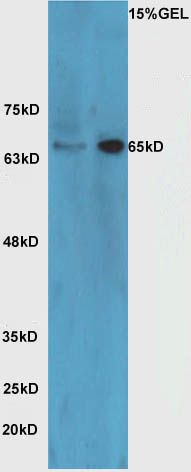
Fig1: Sample: Brain (Mouse) Lysate at 40 ug Raji Cell (Human) Lysate at 40 ug Primary: Anti-HRG beta 1 (ER1910-80) at 1/300 dilution Secondary: HRP conjugated Goat-Anti-rabbit IgG (bs-0295G-HRP) at 1/5000 dilution Predicted band size: 70 kD Observed band size: 65 kDApplication
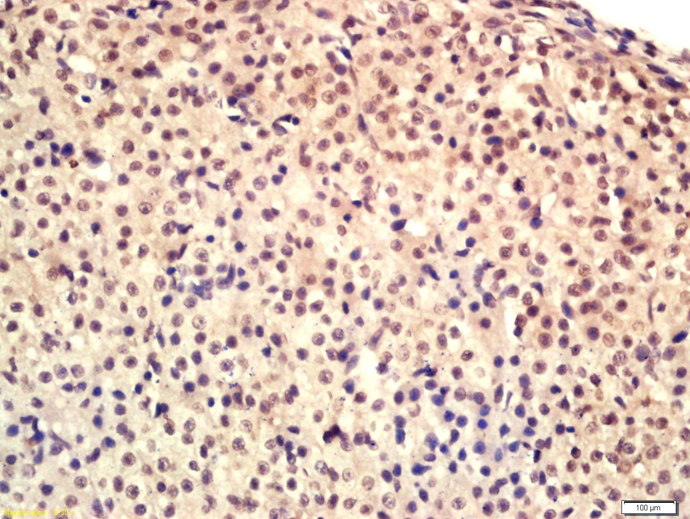
Fig2: Tissue/cell: Mouse ovary tissue; 4% Paraformaldehyde-fixed and paraffin-embedded; Antigen retrieval: citrate buffer ( 0.01M, pH 6.0 ), Boiling bathing for 15min; Block endogenous peroxidase by 3% Hydrogen peroxide for 30min; Blocking buffer (normal goat serum,C-0005) at 37℃ for 20 min; Incubation: Anti-HRG beta 1 Polyclonal Antibody, Unconjugated(ER1910-80) 1:200, overnight at 4℃, followed by conjugation to the secondary antibody(SP-0023) and DAB(C-0010) stainingApplication
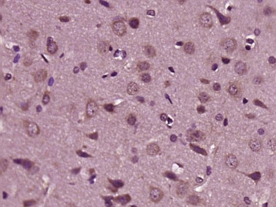
Fig3: Paraformaldehyde-fixed, paraffin embedded (Rat brain); Antigen retrieval by boiling in sodium citrate buffer (pH6.0) for 15min; Block endogenous peroxidase by 3% hydrogen peroxide for 20 minutes; Blocking buffer (normal goat serum) at 37℃ for 30min; Antibody incubation with (HRG beta 1) Polyclonal Antibody, Unconjugated (ER1910-80) at 1:400 overnight at 4℃, followed by operating according to SP Kit(Rabbit) (sp-0023) instructionsand DAB staining.Application
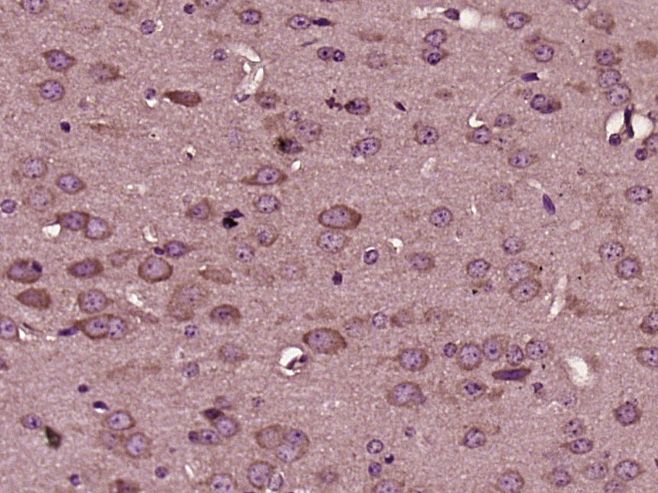
Fig4: Paraformaldehyde-fixed, paraffin embedded (Mouse brain); Antigen retrieval by boiling in sodium citrate buffer (pH6.0) for 15min; Block endogenous peroxidase by 3% hydrogen peroxide for 20 minutes; Blocking buffer (normal goat serum) at 37℃ for 30min; Antibody incubation with (HRG beta 1) Polyclonal Antibody, Unconjugated (ER1910-80) at 1:400 overnight at 4℃, followed by operating according to SP Kit(Rabbit) (sp-0023) instructionsand DAB staining.| Product Name | HRG beta 1 |
|---|---|
| Antibody Type | Primary Antibodies |
| Antigen Alias | Neuregulin 1 GGF2 Acetylcholine receptor inducing activity ARIA Breast cancer cell differentiation factor p45 GGF glial growth factor Heregulin Heregulin beta1 heregulin, alpha (45kD, ERBB2 p185 activator) HGL HRG HRG beta 1A HRG1 HRGA NDF neu differentiation factor NRG1 sensory and motor neuron derived factor SMDF NRG1_HUMAN. |
| Product description | Direct ligand for ERBB3 and ERBB4 tyrosine kinase receptors. Concomitantly recruits ERBB1 and ERBB2 coreceptors, resulting in ligand-stimulated tyrosine phosphorylation and activation of the ERBB receptors. The multiple isoforms perform diverse functions such as inducing growth and differentiation of epithelial, glial, neuronal, and skeletal muscle cells; inducing expression of acetylcholine receptor in synaptic vesicles during the formation of the neuromuscular junction; stimulating lobuloalveolar budding and milk production in the mammary gland and inducing differentiation of mammary tumor cells; stimulating Schwann cell proliferation; implication in the development of the myocardium such as trabeculation of the developing heart. Isoform 10 may play a role in motor and sensory neuron development. |
| Immunogen | KLH conjugated synthetic peptide derived from human Neuregulin 1 65-150/640 |
| Clonality | Polyclonal |
|---|---|
| Isotype | IgG |
| Host Species | Rabbit |
| Tested Applications | WBIHC-PICC/IF |
| WB:1:500-2000: IHC-P:1:400-800: IF:1:200-800: ICC:1:100-500: |
|
| Species Reactivity | HumanMouseRatRabbit |
| Concentration | 1mg/ml |
| Alternative Names | Neuregulin 1
GGF2
Acetylcholine receptor inducing activity
ARIA
Breast cancer cell differentiation factor p45
GGF
glial growth factor
Heregulin
Heregulin beta1
heregulin alpha (45kD ERBB2 p185 activator) HGL HRG HRG beta 1A HRG1 HRGA NDF neu differentiation factor NRG1 sensory and motor neuron derived factor SMDF NRG1_HUMAN. |
|---|---|
| Molecular Weight(MW) | 70 KDa |
| Cellular Localization | Pro-neuregulin-1, membrane-bound isoform: Cell membrane; Single-pass type I membrane protein. Neuregulin-1: Secreted. Isoform 8: Nucleus. Isoform 9: Secreted. Isoform 10: Membrane; Single-pass type I membrane protein. |
| SwissProt ID | Q02297 |
|---|
Application
Fig1: Sample: Brain (Mouse) Lysate at 40 ug Raji Cell (Human) Lysate at 40 ug Primary: Anti-HRG beta 1 (ER1910-80) at 1/300 dilution Secondary: HRP conjugated Goat-Anti-rabbit IgG (bs-0295G-HRP) at 1/5000 dilution Predicted band size: 70 kD Observed band size: 65 kD
Application
Fig2: Tissue/cell: Mouse ovary tissue; 4% Paraformaldehyde-fixed and paraffin-embedded; Antigen retrieval: citrate buffer ( 0.01M, pH 6.0 ), Boiling bathing for 15min; Block endogenous peroxidase by 3% Hydrogen peroxide for 30min; Blocking buffer (normal goat serum,C-0005) at 37℃ for 20 min; Incubation: Anti-HRG beta 1 Polyclonal Antibody, Unconjugated(ER1910-80) 1:200, overnight at 4℃, followed by conjugation to the secondary antibody(SP-0023) and DAB(C-0010) staining
Application
Fig3: Paraformaldehyde-fixed, paraffin embedded (Rat brain); Antigen retrieval by boiling in sodium citrate buffer (pH6.0) for 15min; Block endogenous peroxidase by 3% hydrogen peroxide for 20 minutes; Blocking buffer (normal goat serum) at 37℃ for 30min; Antibody incubation with (HRG beta 1) Polyclonal Antibody, Unconjugated (ER1910-80) at 1:400 overnight at 4℃, followed by operating according to SP Kit(Rabbit) (sp-0023) instructionsand DAB staining.
Application
Fig4: Paraformaldehyde-fixed, paraffin embedded (Mouse brain); Antigen retrieval by boiling in sodium citrate buffer (pH6.0) for 15min; Block endogenous peroxidase by 3% hydrogen peroxide for 20 minutes; Blocking buffer (normal goat serum) at 37℃ for 30min; Antibody incubation with (HRG beta 1) Polyclonal Antibody, Unconjugated (ER1910-80) at 1:400 overnight at 4℃, followed by operating according to SP Kit(Rabbit) (sp-0023) instructionsand DAB staining.| Positive Control | Type I isoforms are the predominant forms expressed in the endocardium. Isoform alpha is expressed in breast, ovary, testis, prostate, heart, skeletal muscle, lung, placenta liver, kidney, salivary gland, small intestine and brain, but not in uterus, stomach, pancreas, and spleen. Isoform 3 is the predominant form in mesenchymal cells and in non-neuronal organs, whereas isoform 6 is the major neuronal form. Isoform 8 is expressed in spinal cord and brain. Isoform 9 is the major form in skeletal muscle cells; in the nervous system it is expressed in spinal cord and brain. Also detected in adult heart, placenta, lung, liver, kidney, and pancreas. Isoform 10 is expressed in nervous system: spinal cord motor neurons, dorsal root ganglion neurons, and brain. Predominant isoform expressed in sensory and motor neurons. Not detected in adult heart, placenta, lung, liver, skeletal muscle, kidney, and pancreas. Not expressed in fetal lung, liver and kidney. Type IV isoforms are brain-specific. |
|---|---|
| Application Notes | WB:1:500-2000: IHC-P:1:400-800: IF:1:200-800: ICC:1:100-500: |
| Form | Liquid |
|---|---|
| Storage Instructions | Store at -20℃ for one year. Avoid repeated freeze/thaw cycles. The lyophilized antibody is stable at room temperature for at least one month and for greater than a year when kept at -20℃. When reconstituted in sterile pH 7.4 0.01M PBS or diluent of antibody the antibody is stable for at least two weeks at 2-4℃. |
| Storage Buffer | 0.01M TBS(pH7.4) with 1% BSA, 0.03% Proclin300 and 50% Glycerol. |


