Application
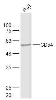
Fig1: Sample: Raji(Human) Cell Lysate at 30 ug Primary: Anti-CD54 (ER1910-98) at 1/1000 dilution Secondary: IRDye800CW Goat Anti-Rabbit IgG at 1/20000 dilution Predicted band size: 56 kD Observed band size: 56 kDApplication
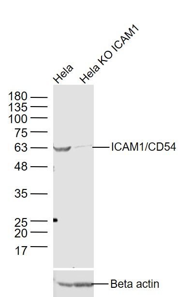
Fig2: Sample: Hela(Human) Cell Lysate at 30 ug Hela KO ICAM1 (Human) Cell Lysate at 30 ug Primary: Anti-ICAM1/CD54 (ER1910-98) at 1/1000 dilution Secondary: IRDye800CW Goat Anti-Rabbit IgG at 1/20000 dilution Predicted band size: 56 kD Observed band size: 56 kDApplication
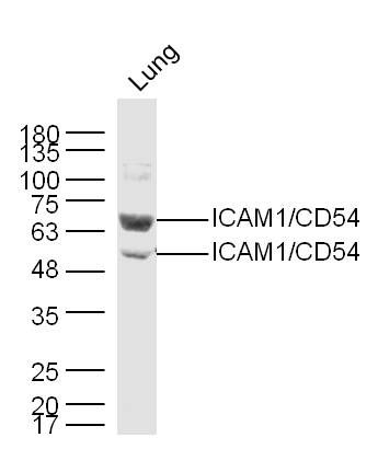
Fig3: Sample: Lung (Mouse) Lysate at 40 ug Primary: Anti-ICAM1 (ER1910-98) at 1/300 dilution Secondary: IRDye800CW Goat Anti-Rabbit IgG at 1/20000 dilution Predicted band size: 56 kD Observed band size: 56/69 kDApplication
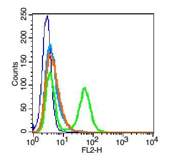
Fig4: Blank control: HUVEC cells(blue). Primary Antibody:Rabbit Anti-CD54 antibody(ER1910-98), Dilution: 1μg in 100 μL 1X PBS containing 0.5% BSA; Isotype Control Antibody: Rabbit IgG(orange) ,used under the same conditions ); Secondary Antibody: Goat anti-rabbit IgG-PE(white blue), Dilution: 1:200 in 1 X PBS containing 0.5% BSA. Protocol The cells were fixed with 2% paraformaldehyde (10 min) .Primary antibody (ER1910-98, 1μg /1x10^6 cells) were incubated for 30 min on the ice, followed by 1 X PBS containing 0.5% BSA + 1 0% goat serum (15 min) to block non-specific protein-protein interactions. Then the Goat Anti-rabbit IgG/PE antibody was added into the blocking buffer mentioned above to react with the primary antibody at 1/200 dilution for 30 min on ice. Acquisition of 20,000 events was performed.Application
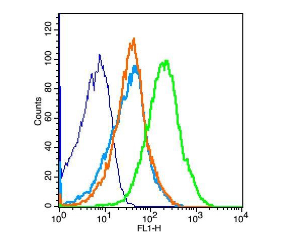
Fig5: Blank control: mouse thymouses(blue) Isotype Control Antibody: Rabbit IgG(orange) ; Secondary Antibody: Goat anti-rabbit IgG-FITC(white blue), Dilution: 1:100 in 1 X PBS containing 0.5% BSA ; Primary Antibody Dilution: 1μg in 100 μL 1X PBS containing 0.5% BSA(green).Application
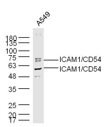
Fig6: Sample: A549(Human) Cell Lysate at 30 ug Primary: Anti-ICAM1 (ER1910-98) at 1/300 dilution Secondary: IRDye800CW Goat Anti-Rabbit IgG at 1/20000 dilution Predicted band size: 56 kD Observed band size: 56/69 kDApplication
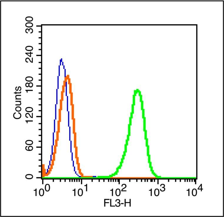
Fig7: Blank control (blue line): A431 cells(blue). Primary Antibody (green line): Rabbit Anti-ICAM1/PE-CY7 Conjugated antibody (ER1910-98-PE-CY7) Dilution: 1μg /10^6 cells; Isotype Control Antibody (orange line): Rabbit IgG-PE-CY7 . Protocol The cells were fixed with 70% ice-cold methanol overnight at 4℃ . The cells were then incubated in 1 X PBS/2%BSA/10% goat serum to block non-specific protein-protein interactions followed by the antibody for 15 min at room temperature. Cells stained with Primary Antibody for 30 min at room temperature.Acquisition of 20,000 events was performed.Application
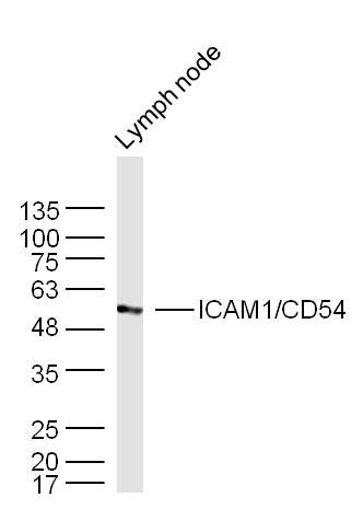
Fig8: Sample: Lymph node (Mouse) Lysate at 40 ug Primary: Anti-ICAM1 (ER1910-98) at 1/300 dilution Secondary: IRDye800CW Goat Anti-Rabbit IgG at 1/20000 dilution Predicted band size: 56 kD Observed band size: 56 kDApplication
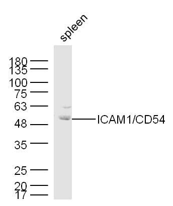
Fig9: Sample: Spleen (Mouse) Lysate at 40 ug Primary: Anti-ICAM1 (ER1910-98) at 1/300 dilution Secondary: IRDye800CW Goat Anti-Rabbit IgG at 1/20000 dilution Predicted band size: 56 kD Observed band size: 56/69 kDWB
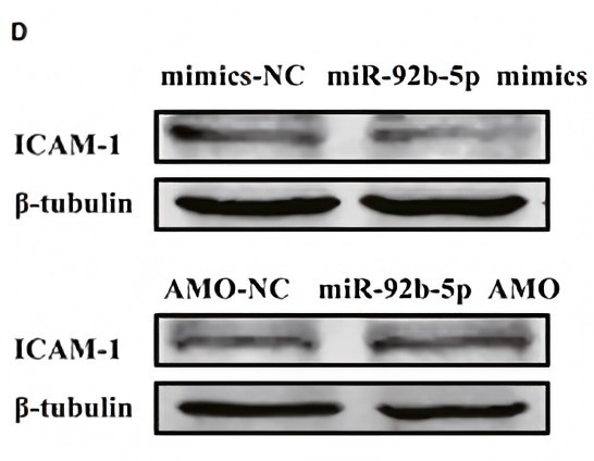
(D) Western blot was performed to detect the protein expression of ICAM-1 after transfection of miR-92b-5p| Product Name | ICAM1/CD54 |
|---|---|
| Antibody Type | Primary Antibodies |
| Product description | In molecular biology, intercellular adhesion molecules (ICAMs) and vascular cell adhesion molecule-1 (VCAM-1) are part of the immunoglobulin superfamily. They are important in inflammation, immune responses and in intracellular signalling events. The ICAM family consists of five members, designated ICAM-1 to ICAM-5. They are known to bind to leucocyte integrins CD11/CD18 such as LFA-1 and Macrophage-1 antigen, during inflammation and in immune responses. In addition, ICAMs may exist in soluble forms in human plasma, due to activation and proteolysis mechanisms at cell surfaces. ICAM proteins are ligands for the leukocyte adhesion protein LFA-1 (integrin alpha-L/beta-2). During leukocyte trans-endothelial migration, ICAM1 engagement promotes the assembly of endothelial apical cups through ARHGEF26/SGEF and RHOG activation. |
| Immunogen | KLH conjugated synthetic peptide derived from human CD54 201-300/537 |
| Clonality | Polyclonal |
|---|---|
| Isotype | IgG |
| Host Species | Rabbit |
| Tested Applications | FCIHC-PWB |
| WB:1:500-2000: IHC-P:1:400-800: FC:1μg/Test: |
|
| Species Reactivity | HumanMouseRat |
| Concentration | 1mg/ml |
| Alternative Names | BB2
CD54 antigen
ICAM 1
ICAM-1
Cell surface glycoprotein P3.58
Human rhinovirus receptor
Intercellular adhesion molecule 1
Major group rhinovirus receptor
MALA2
MyD10
P3.58
Surface antigen of activated B cells
ICAM1_HUMAN
MALA-2
MyD10
intercellular adhesion molecule 1 precursor
intercellular adhesion molecule 1 (CD54) human rhinovirus receptor major group rhinovirus receptor. |
|---|---|
| Molecular Weight(MW) | 56 KDa |
| Cellular Localization | Membrane; Single-pass type I membrane protein. |
| SwissProt ID | P13597 |
|---|
Application
Fig1: Sample: Raji(Human) Cell Lysate at 30 ug Primary: Anti-CD54 (ER1910-98) at 1/1000 dilution Secondary: IRDye800CW Goat Anti-Rabbit IgG at 1/20000 dilution Predicted band size: 56 kD Observed band size: 56 kD
Application
Fig2: Sample: Hela(Human) Cell Lysate at 30 ug Hela KO ICAM1 (Human) Cell Lysate at 30 ug Primary: Anti-ICAM1/CD54 (ER1910-98) at 1/1000 dilution Secondary: IRDye800CW Goat Anti-Rabbit IgG at 1/20000 dilution Predicted band size: 56 kD Observed band size: 56 kD
Application
Fig3: Sample: Lung (Mouse) Lysate at 40 ug Primary: Anti-ICAM1 (ER1910-98) at 1/300 dilution Secondary: IRDye800CW Goat Anti-Rabbit IgG at 1/20000 dilution Predicted band size: 56 kD Observed band size: 56/69 kD
Application
Fig4: Blank control: HUVEC cells(blue). Primary Antibody:Rabbit Anti-CD54 antibody(ER1910-98), Dilution: 1μg in 100 μL 1X PBS containing 0.5% BSA; Isotype Control Antibody: Rabbit IgG(orange) ,used under the same conditions ); Secondary Antibody: Goat anti-rabbit IgG-PE(white blue), Dilution: 1:200 in 1 X PBS containing 0.5% BSA. Protocol The cells were fixed with 2% paraformaldehyde (10 min) .Primary antibody (ER1910-98, 1μg /1x10^6 cells) were incubated for 30 min on the ice, followed by 1 X PBS containing 0.5% BSA + 1 0% goat serum (15 min) to block non-specific protein-protein interactions. Then the Goat Anti-rabbit IgG/PE antibody was added into the blocking buffer mentioned above to react with the primary antibody at 1/200 dilution for 30 min on ice. Acquisition of 20,000 events was performed.
Application
Fig5: Blank control: mouse thymouses(blue) Isotype Control Antibody: Rabbit IgG(orange) ; Secondary Antibody: Goat anti-rabbit IgG-FITC(white blue), Dilution: 1:100 in 1 X PBS containing 0.5% BSA ; Primary Antibody Dilution: 1μg in 100 μL 1X PBS containing 0.5% BSA(green).
Application
Fig6: Sample: A549(Human) Cell Lysate at 30 ug Primary: Anti-ICAM1 (ER1910-98) at 1/300 dilution Secondary: IRDye800CW Goat Anti-Rabbit IgG at 1/20000 dilution Predicted band size: 56 kD Observed band size: 56/69 kD
Application
Fig7: Blank control (blue line): A431 cells(blue). Primary Antibody (green line): Rabbit Anti-ICAM1/PE-CY7 Conjugated antibody (ER1910-98-PE-CY7) Dilution: 1μg /10^6 cells; Isotype Control Antibody (orange line): Rabbit IgG-PE-CY7 . Protocol The cells were fixed with 70% ice-cold methanol overnight at 4℃ . The cells were then incubated in 1 X PBS/2%BSA/10% goat serum to block non-specific protein-protein interactions followed by the antibody for 15 min at room temperature. Cells stained with Primary Antibody for 30 min at room temperature.Acquisition of 20,000 events was performed.
Application
Fig8: Sample: Lymph node (Mouse) Lysate at 40 ug Primary: Anti-ICAM1 (ER1910-98) at 1/300 dilution Secondary: IRDye800CW Goat Anti-Rabbit IgG at 1/20000 dilution Predicted band size: 56 kD Observed band size: 56 kD
Application
Fig9: Sample: Spleen (Mouse) Lysate at 40 ug Primary: Anti-ICAM1 (ER1910-98) at 1/300 dilution Secondary: IRDye800CW Goat Anti-Rabbit IgG at 1/20000 dilution Predicted band size: 56 kD Observed band size: 56/69 kD
WB
(D) Western blot was performed to detect the protein expression of ICAM-1 after transfection of miR-92b-5p| Positive Control | Raji cell, Hela cell, Mouse Lung Lysate, rat brain tissue, HUVEC cells, mouse thymouses, A549 cell, A431 cell, Mouse Lymph node Lysate, Mouse Spleen Lysate. |
|---|---|
| Application Notes | WB:1:500-2000: IHC-P:1:400-800: FC:1μg/Test: |
| Form | Liquid |
|---|---|
| Storage Instructions | Store at -20℃ for one year. Avoid repeated freeze/thaw cycles. The lyophilized antibody is stable at room temperature for at least one month and for greater than a year when kept at -20℃. When reconstituted in sterile pH 7.4 0.01M PBS or diluent of antibody the antibody is stable for at least two weeks at 2-4℃. |
| Storage Buffer | 0.01M TBS(pH7.4) with 1% BSA, 0.03% Proclin300 and 50% Glycerol. |


