Application
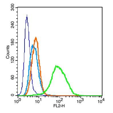
Fig1: Blank control: U937 (blue). Primary Antibody:Rabbit Anti- CD25 antibody(ER1911-34), Dilution: 0.2μg in 100 μL 1X PBS containing 0.5% BSA; Isotype Control Antibody: Rabbit IgG(orange) ,used under the same conditions ); Secondary Antibody: Goat anti-rabbit IgG-PE(white blue), Dilution: 1:200 in 1 X PBS containing 0.5% BSA. Protocol The cells were fixed with 2% paraformaldehyde (10 min) . Primary antibody (ER1911-34, 0.2μg /1x10^6 cells) were incubated for 30 min on the ice, followed by 1 X PBS containing 0.5% BSA + 1 0% goat serum (15 min) to block non-specific protein-protein interactions. Then the Goat Anti-rabbit IgG/PE antibody was added into the blocking buffer mentioned above to react with the primary antibody at 1/200 dilution for 30 min on ice. Acquisition of 20,000 events was performed.Application
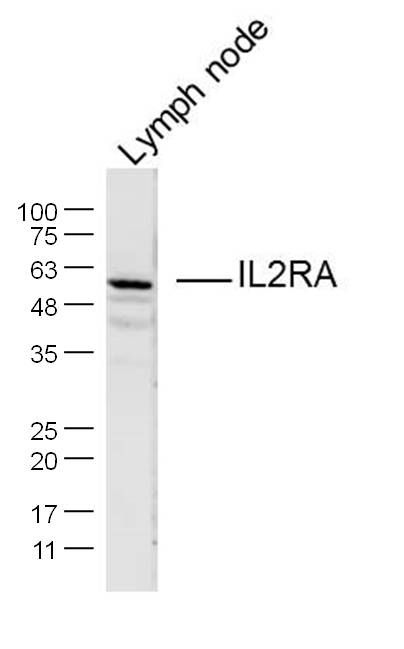
Fig2: Sample: Lymph node (Mouse) Lysate at 40 ug Primary: Anti-IL2RA (ER1911-34) at 1/300 dilution Secondary: IRDye800CW Goat Anti-Rabbit IgG at 1/10000 dilution Predicted band size: 28 kD Observed band size: 55 kDApplication
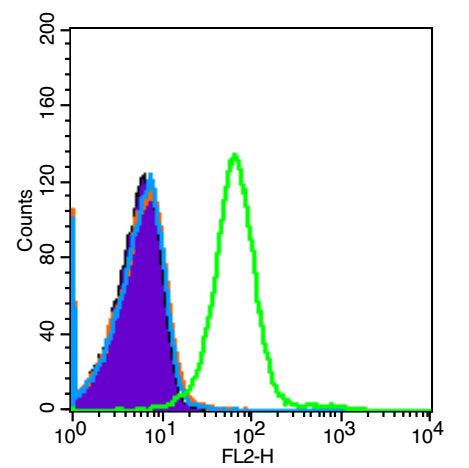
Fig3: Blank control (Black line): Jurkat (Black). Primary Antibody (green line): Rabbit Anti-IL2RA/CD25 antibody (bs-3152R) Dilution: 3μg /10^6 cells; Isotype Control Antibody (orange line): Rabbit IgG . Secondary Antibody (white blue line): Goat anti-rabbit IgG-PE Dilution: 1μg /test. Protocol The cells were fixed with 4% PFA (10min at room temperature)and then were incubated in 5%BSA to block non-specific protein-protein interactions for 15 min at room temperature .Cells stained with Primary Antibody for 30 min at room temperature. The secondary antibody used for 40 min at room temperature. Acquisition of 20,000 events was performed.| Product Name | IL2RA/CD25 |
|---|---|
| Antibody Type | Primary Antibodies |
| Antigen Alias | Interleukin 2 receptor alpha chain CD 25 CD25 CD25 antigen IDDM10 IL 2 RA IL 2 receptor alpha subunit IL2 RA IL-2RA IL2R IL2RA IL2RAC Interleukin 2 Receptor Alpha Interleukin 2 receptor alpha chain Interleukin 2 receptor Ly43 p55 T Cell Growth Factor Receptor TAC Antigen TCGFR IL2RA_MOUSE. |
| Product description | The interleukin 2 (IL2) receptor alpha (IL2RA) and beta (IL2RB) chains, together with the common gamma chain (IL2RG), constitute the high-affinity IL2 receptor. Homodimeric alpha chains (IL2RA) result in low-affinity receptor, while homodimeric beta (IL2RB) chains produce a medium-affinity receptor. Normally an integral-membrane protein, soluble IL2RA has been isolated and determined to result from extracellular proteolysis. Alternately-spliced IL2RA mRNAs have been isolated, but the significance of each is currently unknown. IL2RA is expressed in most B-cell neoplasms, some acute nonlymphocytic leukemias, neuroblastomas, mastocytosis and tumor infiltrating lymphocytes. It functions as the receptor for HTLV-1 and is consequently expressed on neoplastic cells in adult T cell lymphoma/leukemia. Its soluble form, called sIL-2R may be elevated in these diseases and is occasionally used to track disease progression. |
| Immunogen | KLH conjugated synthetic peptide derived from mouse CD25 201-268/268 |
| Clonality | Polyclonal |
|---|---|
| Isotype | IgG |
| Host Species | Rabbit |
| Tested Applications | WBFC |
| WB:1:500-2000: FC:0.2ug /test: |
|
| Species Reactivity | HumanMouseRat |
| Concentration | 1mg/ml |
| Alternative Names | Interleukin 2 receptor alpha chain CD 25 CD25 CD25 antigen IDDM10 IL 2 RA IL 2 receptor alpha subunit IL2 RA IL-2RA IL2R IL2RA IL2RAC Interleukin 2 Receptor Alpha Interleukin 2 receptor alpha chain Interleukin 2 receptor Ly43 p55 T Cell Growth Factor Receptor TAC Antigen TCGFR IL2RA_MOUSE. |
|---|---|
| Molecular Weight(MW) | 28 KDa |
| Cellular Localization | Membrane; Single-pass type I membrane protein. |
| SwissProt ID | P01590 |
|---|
Application
Fig1: Blank control: U937 (blue). Primary Antibody:Rabbit Anti- CD25 antibody(ER1911-34), Dilution: 0.2μg in 100 μL 1X PBS containing 0.5% BSA; Isotype Control Antibody: Rabbit IgG(orange) ,used under the same conditions ); Secondary Antibody: Goat anti-rabbit IgG-PE(white blue), Dilution: 1:200 in 1 X PBS containing 0.5% BSA. Protocol The cells were fixed with 2% paraformaldehyde (10 min) . Primary antibody (ER1911-34, 0.2μg /1x10^6 cells) were incubated for 30 min on the ice, followed by 1 X PBS containing 0.5% BSA + 1 0% goat serum (15 min) to block non-specific protein-protein interactions. Then the Goat Anti-rabbit IgG/PE antibody was added into the blocking buffer mentioned above to react with the primary antibody at 1/200 dilution for 30 min on ice. Acquisition of 20,000 events was performed.
Application
Fig2: Sample: Lymph node (Mouse) Lysate at 40 ug Primary: Anti-IL2RA (ER1911-34) at 1/300 dilution Secondary: IRDye800CW Goat Anti-Rabbit IgG at 1/10000 dilution Predicted band size: 28 kD Observed band size: 55 kD
Application
Fig3: Blank control (Black line): Jurkat (Black). Primary Antibody (green line): Rabbit Anti-IL2RA/CD25 antibody (bs-3152R) Dilution: 3μg /10^6 cells; Isotype Control Antibody (orange line): Rabbit IgG . Secondary Antibody (white blue line): Goat anti-rabbit IgG-PE Dilution: 1μg /test. Protocol The cells were fixed with 4% PFA (10min at room temperature)and then were incubated in 5%BSA to block non-specific protein-protein interactions for 15 min at room temperature .Cells stained with Primary Antibody for 30 min at room temperature. The secondary antibody used for 40 min at room temperature. Acquisition of 20,000 events was performed.| Positive Control | U937 cell, Lymph node (Mouse) Lysate, Jurkat cell. |
|---|---|
| Application Notes | WB:1:500-2000: FC:0.2ug /test: |
| Form | Liquid |
|---|---|
| Storage Instructions | Store at -20℃ for one year. Avoid repeated freeze/thaw cycles. The lyophilized antibody is stable at room temperature for at least one month and for greater than a year when kept at -20℃. When reconstituted in sterile pH 7.4 0.01M PBS or diluent of antibody the antibody is stable for at least two weeks at 2-4℃. |
| Storage Buffer | 0.01M TBS(pH7.4) with 1% BSA, 0.03% Proclin300 and 50% Glycerol. |


