Application
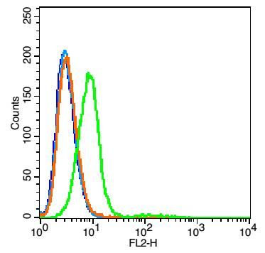
Fig1: Blank control: RSC96 cells(blue). Primary Antibody: Rabbit Anti-Integrin alpha 1 antibody(ER1911-50), Dilution: 5μg in 100 μL 1X PBS containing 0.5% BSA; Isotype Control Antibody: Rabbit IgG (orange) ,used under the same conditions. Secondary Antibody: Goat anti-rabbit IgG-PE(white blue), Dilution: 1:200 in 1 X PBS containing 0.5% BSA.Application
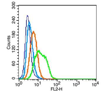
Fig2: Blank control: U937(blue). Primary Antibody: Rabbit Anti- Integrin alpha 1 antibody(ER1911-50), Dilution: 1μg in 100 μL 1X PBS containing 0.5% BSA; Isotype Control Antibody: Rabbit IgG(orange) ,used under the same conditions ); Secondary Antibody: Goat anti-rabbit IgG-PE(white blue), Dilution: 1:200 in 1 X PBS containing 0.5% BSA. Protocol The cells were fixed with 2% paraformaldehyde (10 min). Primary antibody (ER1911-50, 1μg /1x10^6 cells) were incubated for 30 min on the ice, followed by 1 X PBS containing 0.5% BSA + 1 0% goat serum (15 min) to block non-specific protein-protein interactions. Then the Goat Anti-rabbit IgG/PE antibody was added into the blocking buffer mentioned above to react with the primary antibody at 1/200 dilution for 30 min on ice. Acquisition of 20,000 events was performed.Application
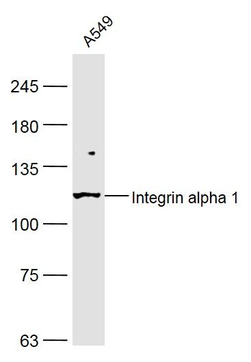
Fig3: Sample: A549 (Human) Cell Lysate at 40 ug Primary: Anti-Integrin alpha 1 (ER1911-50) at 1/300 dilution Secondary: IRDye800CW Goat Anti-Rabbit IgG at 1/20000 dilution Predicted band size: 128 kD Observed band size: 128 kDApplication
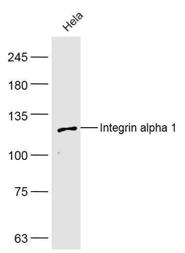
Fig4: Sample: Hela (Human) Cell Lysate at 40 ug Primary: Anti-Integrin alpha 1 (ER1911-50) at 1/300 dilution Secondary: IRDye800CW Goat Anti-Rabbit IgG at 1/20000 dilution Predicted band size: 128 kD Observed band size: 128 kD| Product Name | Integrin alpha 1 |
|---|---|
| Antibody Type | Primary Antibodies |
| Antigen Alias | CD 49a CD49a CD49 antigen-like family member A CD49a CD49a antigen Integrin alpha-1 ITA1_HUMAN Itga 1 ITGA1 Laminin and collagen receptor Very late activation protein 1 VLA 1 VLA-1 VLA1. |
| Product description | Integrin alpha-1/beta-1 is a receptor for laminin and collagen. It recognizes the proline-hydroxylated sequence G-F-P-G-E-R in collagen. |
| Immunogen | KLH conjugated synthetic peptide derived from human Integrin alpha 1 1001-1179/1179 |
| Clonality | Polyclonal |
|---|---|
| Isotype | IgG |
| Host Species | Rabbit |
| Tested Applications | WBFC |
| WB:1:500-2000: FC:1μg /test: |
|
| Species Reactivity | HumanMouseRatCowHorse |
| Concentration | 1mg/ml |
| Alternative Names | CD 49a CD49a CD49 antigen-like family member A CD49a CD49a antigen Integrin alpha-1 ITA1_HUMAN Itga 1 ITGA1 Laminin and collagen receptor Very late activation protein 1 VLA 1 VLA-1 VLA1. |
|---|---|
| Molecular Weight(MW) | 128 KDa |
| Cellular Localization | Membrane; Single-pass type I membrane protein. |
| SwissProt ID | P56199 |
|---|
Application
Fig1: Blank control: RSC96 cells(blue). Primary Antibody: Rabbit Anti-Integrin alpha 1 antibody(ER1911-50), Dilution: 5μg in 100 μL 1X PBS containing 0.5% BSA; Isotype Control Antibody: Rabbit IgG (orange) ,used under the same conditions. Secondary Antibody: Goat anti-rabbit IgG-PE(white blue), Dilution: 1:200 in 1 X PBS containing 0.5% BSA.
Application
Fig2: Blank control: U937(blue). Primary Antibody: Rabbit Anti- Integrin alpha 1 antibody(ER1911-50), Dilution: 1μg in 100 μL 1X PBS containing 0.5% BSA; Isotype Control Antibody: Rabbit IgG(orange) ,used under the same conditions ); Secondary Antibody: Goat anti-rabbit IgG-PE(white blue), Dilution: 1:200 in 1 X PBS containing 0.5% BSA. Protocol The cells were fixed with 2% paraformaldehyde (10 min). Primary antibody (ER1911-50, 1μg /1x10^6 cells) were incubated for 30 min on the ice, followed by 1 X PBS containing 0.5% BSA + 1 0% goat serum (15 min) to block non-specific protein-protein interactions. Then the Goat Anti-rabbit IgG/PE antibody was added into the blocking buffer mentioned above to react with the primary antibody at 1/200 dilution for 30 min on ice. Acquisition of 20,000 events was performed.
Application
Fig3: Sample: A549 (Human) Cell Lysate at 40 ug Primary: Anti-Integrin alpha 1 (ER1911-50) at 1/300 dilution Secondary: IRDye800CW Goat Anti-Rabbit IgG at 1/20000 dilution Predicted band size: 128 kD Observed band size: 128 kD
Application
Fig4: Sample: Hela (Human) Cell Lysate at 40 ug Primary: Anti-Integrin alpha 1 (ER1911-50) at 1/300 dilution Secondary: IRDye800CW Goat Anti-Rabbit IgG at 1/20000 dilution Predicted band size: 128 kD Observed band size: 128 kD| Application Notes | WB:1:500-2000: FC:1μg /test: |
|---|
| Form | Liquid |
|---|---|
| Storage Instructions | Store at -20℃ for one year. Avoid repeated freeze/thaw cycles. The lyophilized antibody is stable at room temperature for at least one month and for greater than a year when kept at -20℃. When reconstituted in sterile pH 7.4 0.01M PBS or diluent of antibody the antibody is stable for at least two weeks at 2-4℃. |
| Storage Buffer | 0.01M TBS(pH7.4) with 1% BSA, 0.03% Proclin300 and 50% Glycerol. |


