Application
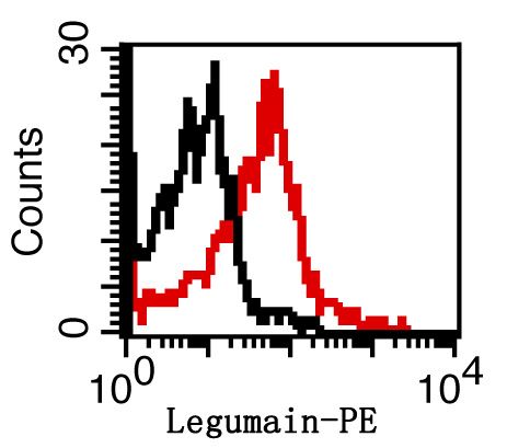
Fig1: Overlay histogram showing Mouse spleen cells stained with ER1912-26-PE (red line). The cells were fixed with 1% paraformaldehyde (10 min).The cells were then incubated with the antibody (ER1912-26-PE, 2ug/1x106cells) for 30 min at 22-25℃. Isotype control antibody (black line) was rabbit IgG (2ug/1x106cells) used under the same conditions. Acquisition of events were used for analysis.Application
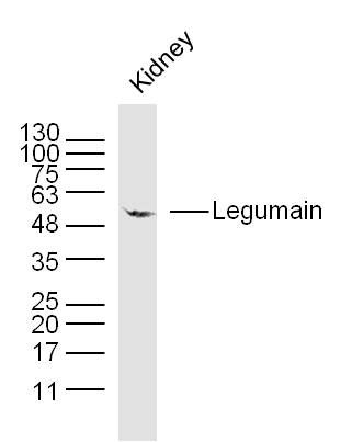
Fig2: Sample: Kidney (Mouse) Lysate at 40 ug Primary: Anti- Legumain (ER1912-26) at 1/300 dilution Secondary: IRDye800CW Goat Anti-Rabbit IgG at 1/20000 dilution Predicted band size: 46 kD Observed band size: 55 kDApplication
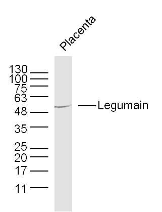
Fig3: Sample: Placenta (Mouse) Lysate at 40 ug Primary: Anti-Legumain (ER1912-26) at 1/300 dilution Secondary: IRDye800CW Goat Anti-Rabbit IgG at 1/20000 dilution Predicted band size: 46 kD Observed band size: 55 kDApplication
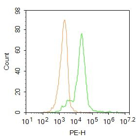
Fig4: Blank control: HL60. Primary Antibody (green line): Rabbit Anti-Legumain antibody (ER1912-26) Dilution: 3μg /10^6 cells; Isotype Control Antibody (orange line): Rabbit IgG . Secondary Antibody : Goat anti-rabbit IgG-PE Dilution: 1μg /test. Protocol The cells were fixed with 4% PFA (10min at room temperature)and then permeabilized with PBST for 20 min at room temperature. The cells were then incubated in 5%BSA to block non-specific protein-protein interactions for 30 min at at room temperature .Cells stained with Primary Antibody for 30 min at room temperature. The secondary antibody used for 40 min at room temperature. Acquisition of 20,000 events was performed.| Product Name | Legumain |
|---|---|
| Antibody Type | Primary Antibodies |
| Antigen Alias | AEP Asparaginyl endopeptidase EC 3.4.22.34 LGMN LGMN1 Protease, cysteine 1 protease, cysteine, 1 (legumain) PRSC1 LGMN_HUMAN. |
| Product description | Has a strict specificity for hydrolysis of asparaginyl bonds. Can also cleave aspartyl bonds slowly, especially under acidic conditions. May be involved in the processing of proteins for MHC class II antigen presentation in the lysosomal/endosomal system. |
| Immunogen | KLH conjugated synthetic peptide derived from human Legumain 201-300/433 |
| Clonality | Polyclonal |
|---|---|
| Isotype | IgG |
| Host Species | Rabbit |
| Tested Applications | WBFC |
| WB:1:500-2000: FC:3ug/Test: |
|
| Species Reactivity | HumanMouseRatDogCowHorse |
| Concentration | 1mg/ml |
| Alternative Names | AEP
Asparaginyl endopeptidase
EC 3.4.22.34
LGMN
LGMN1
Protease cysteine 1 protease cysteine 1 (legumain) PRSC1 LGMN_HUMAN. |
|---|---|
| Molecular Weight(MW) | 46 KDa |
| Cellular Localization | Lysosome (By similarity). |
| SwissProt ID | Q99538 |
|---|
Application
Fig1: Overlay histogram showing Mouse spleen cells stained with ER1912-26-PE (red line). The cells were fixed with 1% paraformaldehyde (10 min).The cells were then incubated with the antibody (ER1912-26-PE, 2ug/1x106cells) for 30 min at 22-25℃. Isotype control antibody (black line) was rabbit IgG (2ug/1x106cells) used under the same conditions. Acquisition of events were used for analysis.
Application
Fig2: Sample: Kidney (Mouse) Lysate at 40 ug Primary: Anti- Legumain (ER1912-26) at 1/300 dilution Secondary: IRDye800CW Goat Anti-Rabbit IgG at 1/20000 dilution Predicted band size: 46 kD Observed band size: 55 kD
Application
Fig3: Sample: Placenta (Mouse) Lysate at 40 ug Primary: Anti-Legumain (ER1912-26) at 1/300 dilution Secondary: IRDye800CW Goat Anti-Rabbit IgG at 1/20000 dilution Predicted band size: 46 kD Observed band size: 55 kD
Application
Fig4: Blank control: HL60. Primary Antibody (green line): Rabbit Anti-Legumain antibody (ER1912-26) Dilution: 3μg /10^6 cells; Isotype Control Antibody (orange line): Rabbit IgG . Secondary Antibody : Goat anti-rabbit IgG-PE Dilution: 1μg /test. Protocol The cells were fixed with 4% PFA (10min at room temperature)and then permeabilized with PBST for 20 min at room temperature. The cells were then incubated in 5%BSA to block non-specific protein-protein interactions for 30 min at at room temperature .Cells stained with Primary Antibody for 30 min at room temperature. The secondary antibody used for 40 min at room temperature. Acquisition of 20,000 events was performed.| Positive Control | Ubiquitous. Particularly abundant in kidney, heart and placenta. |
|---|---|
| Application Notes | WB:1:500-2000: FC:3ug/Test: |
| Form | Liquid |
|---|---|
| Storage Instructions | Store at -20℃ for one year. Avoid repeated freeze/thaw cycles. The lyophilized antibody is stable at room temperature for at least one month and for greater than a year when kept at -20℃. When reconstituted in sterile pH 7.4 0.01M PBS or diluent of antibody the antibody is stable for at least two weeks at 2-4℃. |
| Storage Buffer | 0.01M TBS(pH7.4) with 1% BSA, 0.03% Proclin300 and 50% Glycerol. |


