Application
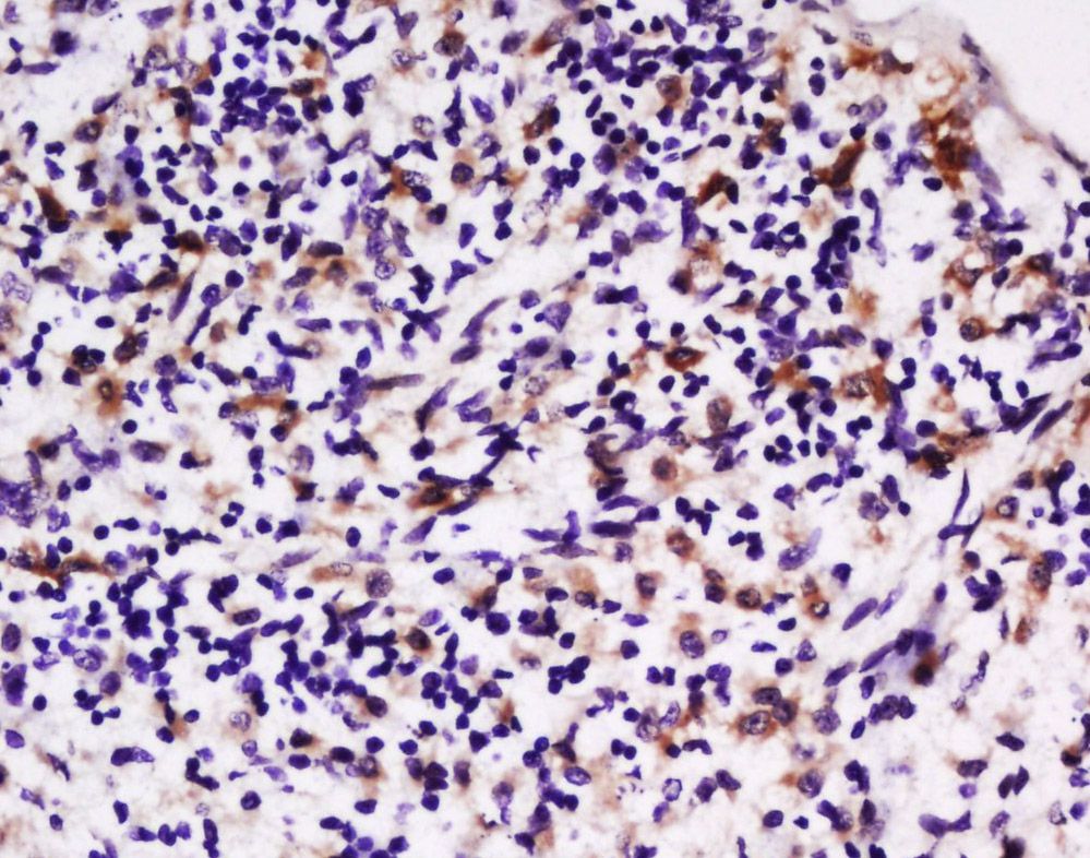
Fig1: Tissue/cell: rat spleen tissue; 4% Paraformaldehyde-fixed and paraffin-embedded; Antigen retrieval: citrate buffer ( 0.01M, pH 6.0 ), Boiling bathing for 15min; Block endogenous peroxidase by 3% Hydrogen peroxide for 30min; Blocking buffer (normal goat serum,C-0005) at 37℃ for 20 min; Incubation: Anti-LPA2 Polyclonal Antibody, Unconjugated(ER1912-45) 1:100, overnight at 4℃, followed by conjugation to the secondary antibody(SP-0023) and DAB(C-0010) stainingApplication
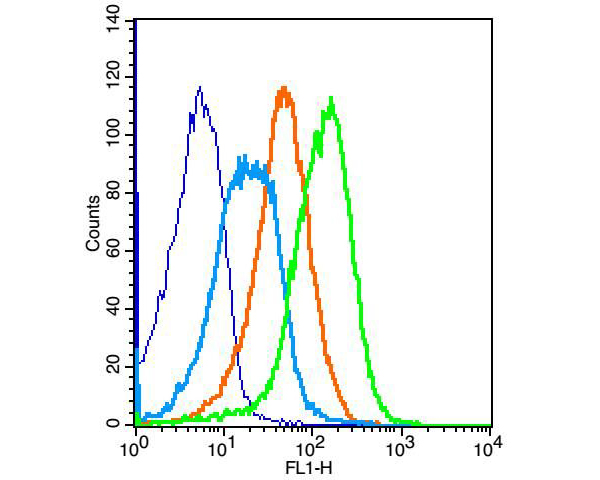
Fig2: Positive control: (mo)Splenocytes(2% Paraformaldehyde-fixed ) Isotype Control Antibody: Rabbit IgG,Dilution: 1μg in 100 μl 1 X PBS containing 0.5% BSA Secondary Antibody: Goat anti-rabbit IgG-FITC,Dilution: 1:200 in 1 X PBS containing 0.5% BSA Primary Antibody: rabbit Anti-LPA2 (ER1912-45),Dilution: 1μg in 100 μl 1X PBS containing 0.5% BSAApplication
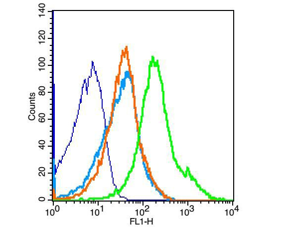
Fig3: Blank control: mouse thymouses(blue) Isotype Control Antibody: Rabbit IgG(orange) ; Secondary Antibody: Goat anti-rabbit IgG-FITC(white blue), Dilution: 1:100 in 1 X PBS containing 0.5% BSA ; Primary Antibody Dilution: 1μl in 100 μL1X PBS containing 0.5% BSA(green).Application
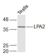
Fig4: Sample: Testis (Rat) Lysate at 40 ug Primary: Anti-LPA2 (ER1912-45) at 1/1000 dilution Secondary: IRDye800CW Goat Anti-Rabbit IgG at 1/20000 dilution Predicted band size: 39 kD Observed band size: 39 kD| Product Name | LPA2 |
|---|---|
| Antibody Type | Primary Antibodies |
| Antigen Alias | Endothelial Cell Differentiation Gene 4 Endothelial differentiation lysophosphatidic acid G protein coupled receptor 4 IPA2 LPA receptor 2 LPA receptor EDG4 LPA2 LPAR2 Lysophosphatidic acid receptor Edg4 Edg4 LPAR2_HUMAN. |
| Product description | Receptor for lysophosphatidic acid (LPA), a mediator of diverse cellular activities. Seems to be coupled to the G(i)/G(o), G(12)/G(13), and G(q) families of heteromeric G proteins. Plays a key role in phospholipase C-beta (PLC-beta) signaling pathway. Stimulates phospholipase C (PLC) activity in a manner that is independent of RALA activation. |
| Immunogen | KLH conjugated synthetic peptide derived from human LPA2 1-100/351 |
| Clonality | Polyclonal |
|---|---|
| Isotype | IgG |
| Host Species | Rabbit |
| Tested Applications | WBIHC-PFC |
| WB:1:500-2000: IHC-P:1:400-800: FC:1μg/Test: |
|
| Species Reactivity | HumanMouseRatDogPigCowHorseRabbitSheep |
| Concentration | 1mg/ml |
| Alternative Names | Endothelial Cell Differentiation Gene 4 Endothelial differentiation lysophosphatidic acid G protein coupled receptor 4 IPA2 LPA receptor 2 LPA receptor EDG4 LPA2 LPAR2 Lysophosphatidic acid receptor Edg4 Edg4 LPAR2_HUMAN. |
|---|---|
| Molecular Weight(MW) | 39 KDa |
| Cellular Localization | Cell surface. Cell membrane; Multi-pass membrane protein. Note=Prior to LPA treatment found predominantly at the cell surface but in the presence of LPA co-localizes with RALA in the endocytic vesicles. |
| SwissProt ID | Q9HBW0 |
|---|
Application
Fig1: Tissue/cell: rat spleen tissue; 4% Paraformaldehyde-fixed and paraffin-embedded; Antigen retrieval: citrate buffer ( 0.01M, pH 6.0 ), Boiling bathing for 15min; Block endogenous peroxidase by 3% Hydrogen peroxide for 30min; Blocking buffer (normal goat serum,C-0005) at 37℃ for 20 min; Incubation: Anti-LPA2 Polyclonal Antibody, Unconjugated(ER1912-45) 1:100, overnight at 4℃, followed by conjugation to the secondary antibody(SP-0023) and DAB(C-0010) staining
Application
Fig2: Positive control: (mo)Splenocytes(2% Paraformaldehyde-fixed ) Isotype Control Antibody: Rabbit IgG,Dilution: 1μg in 100 μl 1 X PBS containing 0.5% BSA Secondary Antibody: Goat anti-rabbit IgG-FITC,Dilution: 1:200 in 1 X PBS containing 0.5% BSA Primary Antibody: rabbit Anti-LPA2 (ER1912-45),Dilution: 1μg in 100 μl 1X PBS containing 0.5% BSA
Application
Fig3: Blank control: mouse thymouses(blue) Isotype Control Antibody: Rabbit IgG(orange) ; Secondary Antibody: Goat anti-rabbit IgG-FITC(white blue), Dilution: 1:100 in 1 X PBS containing 0.5% BSA ; Primary Antibody Dilution: 1μl in 100 μL1X PBS containing 0.5% BSA(green).
Application
Fig4: Sample: Testis (Rat) Lysate at 40 ug Primary: Anti-LPA2 (ER1912-45) at 1/1000 dilution Secondary: IRDye800CW Goat Anti-Rabbit IgG at 1/20000 dilution Predicted band size: 39 kD Observed band size: 39 kD| Positive Control | Expressed most abundantly in testes and peripheral blood leukocytes with less expression in pancreas, spleen, thymus and prostate. Little or no expression in heart, brain, placenta, lung, liver, skeletal muscle, kidney, ovary, small intestine, or colon. |
|---|---|
| Application Notes | WB:1:500-2000: IHC-P:1:400-800: FC:1μg/Test: |
| Form | Liquid |
|---|---|
| Storage Instructions | Store at -20℃ for one year. Avoid repeated freeze/thaw cycles. The lyophilized antibody is stable at room temperature for at least one month and for greater than a year when kept at -20℃. When reconstituted in sterile pH 7.4 0.01M PBS or diluent of antibody the antibody is stable for at least two weeks at 2-4℃. |
| Storage Buffer | 0.01M TBS(pH7.4) with 1% BSA, 0.03% Proclin300 and 50% Glycerol. |


