Application
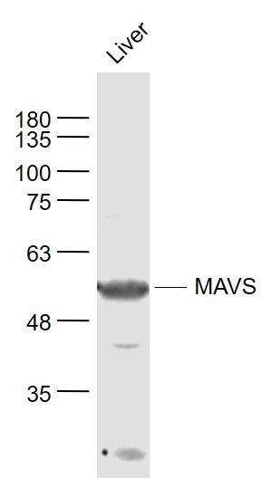
Fig1: Sample: Liver (Mouse) Lysate at 40 ug Primary: Anti- MAVS (ER1912-80) at 1/1000 dilution Secondary: IRDye800CW Goat Anti-Rabbit IgG at 1/20000 dilution Predicted band size: 56 kD Observed band size: 56 kD| Product Name | MAVS |
|---|---|
| Antibody Type | Primary Antibodies |
| Antigen Alias | CARD adapter inducing interferon beta Cardif Interferon beta promoter stimulator protein 1 Interferon-beta promoter stimulator protein 1 Ips 1 Ips1 KIAA1271 Mitochondrial anti viral signaling protein Mitochondrial Antiviral Signaling Mitochondrial antiviral signaling protein Putative NF kappa B activating protein 031N Virus induced signaling adapter DKFZp547C224 DKFZp666M015 FLJ27482 FLJ31698 FLJ35386 FLJ38051 CARDIF FLJ41962 IPS-1 IPS1 MGC3260 VISA MAVS_HUMAN. |
| Product description | Required for innate immune defense against viruses. Acts downstream of DDX58 and IFIH1/MDA5, which detect intracellular dsRNA produced during viral replication, to coordinate pathways leading to the activation of NF-kappa-B, IRF3 and IRF7, and to the subsequent induction of antiviral cytokines such as IFN-beta and RANTES (CCL5). May activate the same pathways following detection of extracellular dsRNA by TLR3. May protect cells from apoptosis. |
| Immunogen | KLH conjugated synthetic peptide derived from human MAVS N terminus 3-100/540 |
| Clonality | Polyclonal |
|---|---|
| Isotype | IgG |
| Host Species | Rabbit |
| Tested Applications | WB |
| WB:1:500-2000: |
|
| Species Reactivity | HumanMouseRatCow |
| Concentration | 1mg/ml |
| Alternative Names | CARD adapter inducing interferon beta Cardif Interferon beta promoter stimulator protein 1 Interferon-beta promoter stimulator protein 1 Ips 1 Ips1 KIAA1271 Mitochondrial anti viral signaling protein Mitochondrial Antiviral Signaling Mitochondrial antiviral signaling protein Putative NF kappa B activating protein 031N Virus induced signaling adapter DKFZp547C224 DKFZp666M015 FLJ27482 FLJ31698 FLJ35386 FLJ38051 CARDIF FLJ41962 IPS-1 IPS1 MGC3260 VISA MAVS_HUMAN. |
|---|---|
| Molecular Weight(MW) | 56 KDa |
| Cellular Localization | Mitochondrion outer membrane. |
| SwissProt ID | Q7Z434 |
|---|
Application
Fig1: Sample: Liver (Mouse) Lysate at 40 ug Primary: Anti- MAVS (ER1912-80) at 1/1000 dilution Secondary: IRDye800CW Goat Anti-Rabbit IgG at 1/20000 dilution Predicted band size: 56 kD Observed band size: 56 kD| Positive Control | Present in T-cells, monocytes, epithelial cells and hepatocytes (at protein level). Ubiquitously expressed, with highest levels in heart, skeletal muscle, liver, placenta and peripheral blood leukocytes. |
|---|---|
| Application Notes | WB:1:500-2000: |
| Form | Liquid |
|---|---|
| Storage Instructions | Store at -20℃ for one year. Avoid repeated freeze/thaw cycles. The lyophilized antibody is stable at room temperature for at least one month and for greater than a year when kept at -20℃. When reconstituted in sterile pH 7.4 0.01M PBS or diluent of antibody the antibody is stable for at least two weeks at 2-4℃. |
| Storage Buffer | 0.01M TBS(pH7.4) with 1% BSA, 0.03% Proclin300 and 50% Glycerol. |


