Application
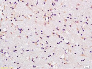
Fig1: Paraformaldehyde-fixed, paraffin embedded (rat brain); Antigen retrieval by boiling in sodium citrate buffer (pH6.0) for 15min; Block endogenous peroxidase by 3% hydrogen peroxide for 20 minutes; Blocking buffer (normal goat serum) at 37℃ for 30min; Antibody incubation with (MCT1) Polyclonal Antibody, Unconjugated (ER1912-87) at 1:400 overnight at 4℃, followed by operating according to SP Kit(Rabbit) (sp-0023) instructionsand DAB staining.Application
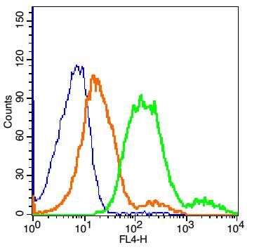
Fig2: Blank control: MCF7 Cells(blue). Primary Antibody: Rabbit Anti-MCT1/AF647 Conjugated antibody (ER1912-87-AF647), Dilution: 1μg in 100 μL 1X PBS containing 0.5% BSA; Isotype Control Antibody: Rabbit IgG/FITC(orange) ,used under the same conditions.Application
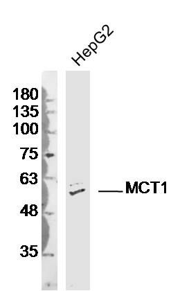
Fig3: Sample:HepG2 (Human)cell Lysate at 40 ug Primary: Anti-MCT1(ER1912-87)at 1/300 dilution Secondary: IRDye800CW Goat Anti-RabbitIgG at 1/20000 dilution Predicted band size: 55kD Observed band size: 55kDWB
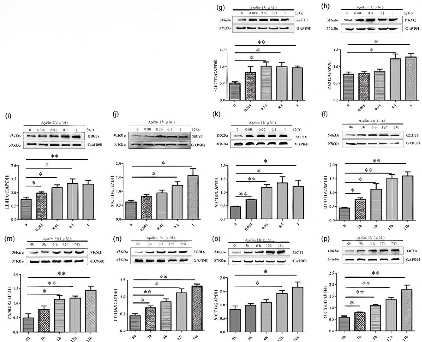
(g–p) Results from western blot analysis showed that apelin-13 promoted the expression of GLUT1, PKM2, LDHA, MCT1, and MCT4 in dose- and time-dependent manners for 24 hr (data were presented as mean ± SEM, n = 3–4; *p < 0.05, **p < 0.01, and ***p < 0.001 vs. control).WB
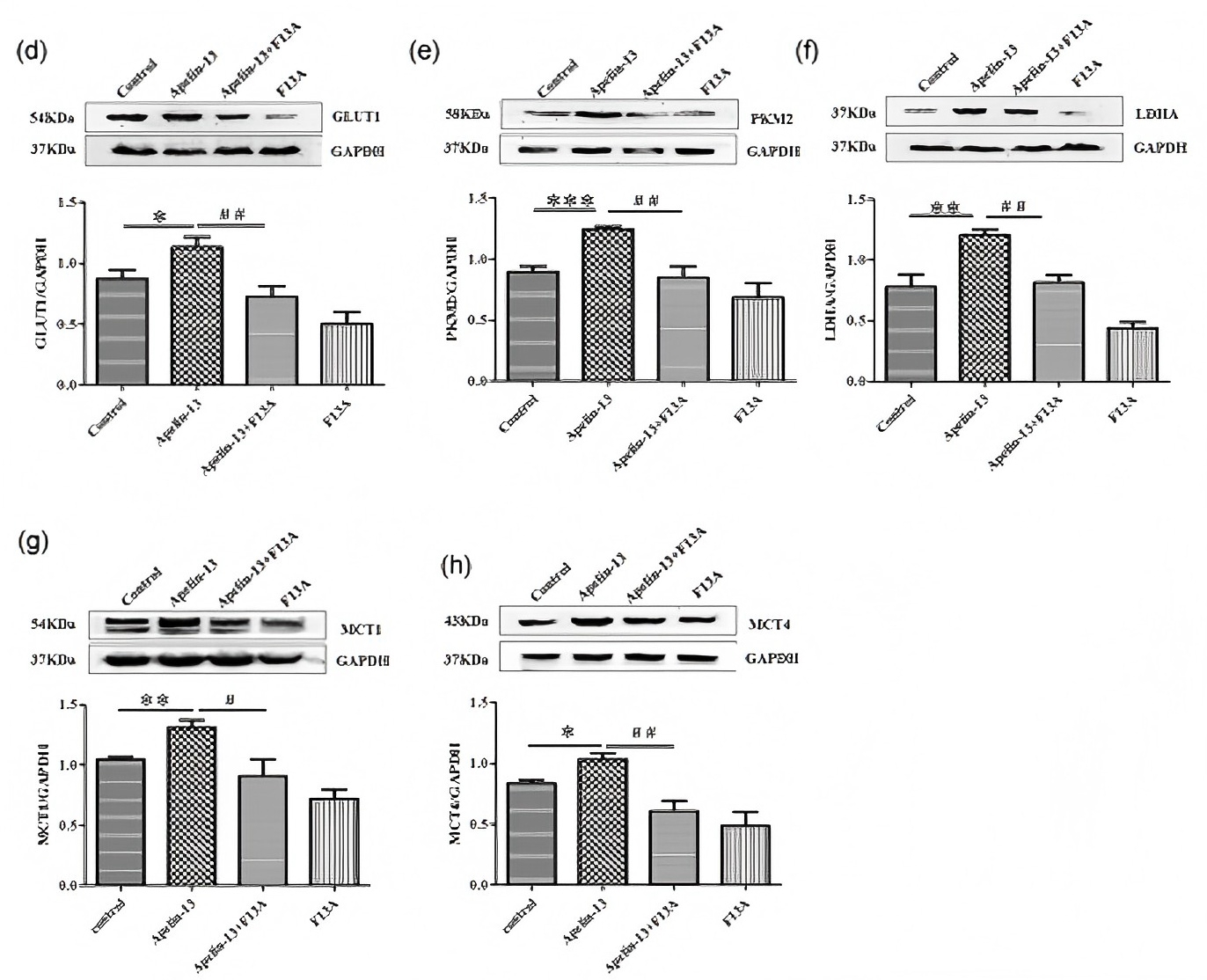
(d–h) Western blot analysis showed that apelin-13 promoted the expression of GLUT1, PKM2, LDHA, MCT1, and MCT4, however, F13A reversed apelin-13-induced GLUT1, PKM2, LDHA, MCT1, and MCT4 expression (data were presented as mean ± SEM, n = 4–5; *p < 0.05, **p < 0.01, and ***p < 0.001 vs.| Product Name | MCT1 |
|---|---|
| Antibody Type | Primary Antibodies |
| Product description | Proton-coupled monocarboxylate transporter. Catalyzes the rapid transport across the plasma membrane of many monocarboxylates such as lactate, pyruvate, branched-chain oxo acids derived from leucine, valine and isoleucine, and the ketone bodies acetoacetate, beta-hydroxybutyrate and acetate. Depending on the tissue and on cicumstances, mediates the import or export of lactic acid and ketone bodies. Required for normal nutrient assimilation, increase of white adipose tissue and body weight gain when on a high-fat diet. Plays a role in cellular responses to a high-fat diet by modulating the cellular levels of lactate and pyruvate, small molecules that contribute to the regulation of central metabolic pathways and insulin secretion, with concomitant effects on plasma insulin levels and blood glucose homeostasis. |
| Immunogen | KLH conjugated synthetic peptide derived from human MCT1 251-350/500 |
| Clonality | Polyclonal |
|---|---|
| Isotype | IgG |
| Host Species | Rabbit |
| Tested Applications | FCIHC-PWB |
| WB:1:500-2000: IHC-P:1:400-800: FC:1μg/Test: |
|
| Species Reactivity | CowDogGuinea PigHorseHumanMousePigRabbitRat |
| Concentration | 1mg/ml |
| Alternative Names | monocarboxylate transporter 1 Malignant T cell amplified sequence 1 MCT 1 MCT1 MCTS 1 MCTS1 Oncogene MCT 1 Oncogene MCT1 SLC16A1 FLJ36745 HHF7 MCT MGC44475 monocarboxylate transporter Monocarboxylate transporter isoform 1 Monocarboxylic acid transporter 1 SLC16A1 SLC16A1 protein Solute carrier family 16 (monocarboxylic acid transporters) member 1 Solute carrier family 16 member 1 (monocarboxylic acid transporter 1) Solute carrier family 16 member 1 MOT1_HUMAN. |
|---|---|
| Molecular Weight(MW) | 55 KDa |
| Cellular Localization | Cell membrane; Multi-pass membrane protein. |
| SwissProt ID | P53985 |
|---|
Application
Fig1: Paraformaldehyde-fixed, paraffin embedded (rat brain); Antigen retrieval by boiling in sodium citrate buffer (pH6.0) for 15min; Block endogenous peroxidase by 3% hydrogen peroxide for 20 minutes; Blocking buffer (normal goat serum) at 37℃ for 30min; Antibody incubation with (MCT1) Polyclonal Antibody, Unconjugated (ER1912-87) at 1:400 overnight at 4℃, followed by operating according to SP Kit(Rabbit) (sp-0023) instructionsand DAB staining.
Application
Fig2: Blank control: MCF7 Cells(blue). Primary Antibody: Rabbit Anti-MCT1/AF647 Conjugated antibody (ER1912-87-AF647), Dilution: 1μg in 100 μL 1X PBS containing 0.5% BSA; Isotype Control Antibody: Rabbit IgG/FITC(orange) ,used under the same conditions.
Application
Fig3: Sample:HepG2 (Human)cell Lysate at 40 ug Primary: Anti-MCT1(ER1912-87)at 1/300 dilution Secondary: IRDye800CW Goat Anti-RabbitIgG at 1/20000 dilution Predicted band size: 55kD Observed band size: 55kD
WB
(g–p) Results from western blot analysis showed that apelin-13 promoted the expression of GLUT1, PKM2, LDHA, MCT1, and MCT4 in dose- and time-dependent manners for 24 hr (data were presented as mean ± SEM, n = 3–4; *p < 0.05, **p < 0.01, and ***p < 0.001 vs. control).
WB
(d–h) Western blot analysis showed that apelin-13 promoted the expression of GLUT1, PKM2, LDHA, MCT1, and MCT4, however, F13A reversed apelin-13-induced GLUT1, PKM2, LDHA, MCT1, and MCT4 expression (data were presented as mean ± SEM, n = 4–5; *p < 0.05, **p < 0.01, and ***p < 0.001 vs.| Positive Control | Widely expressed in normal and in cancer cells. |
|---|---|
| Application Notes | WB:1:500-2000: IHC-P:1:400-800: FC:1μg/Test: |
| Form | Liquid |
|---|---|
| Storage Instructions | Store at -20℃ for one year. Avoid repeated freeze/thaw cycles. The lyophilized antibody is stable at room temperature for at least one month and for greater than a year when kept at -20℃. When reconstituted in sterile pH 7.4 0.01M PBS or diluent of antibody the antibody is stable for at least two weeks at 2-4℃. |
| Storage Buffer | 0.01M TBS(pH7.4) with 1% BSA, 0.03% Proclin300 and 50% Glycerol. |


