Application
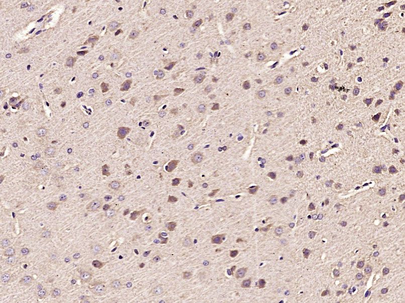
Fig1: Paraformaldehyde-fixed, paraffin embedded (Rat brain); Antigen retrieval by microwave in sodium citrate buffer (pH6.0) ; Block endogenous peroxidase by 3% hydrogen peroxide for 30 minutes; Blocking buffer (3% BSA) at RT for 30min; Antibody incubation with (MDH1) Polyclonal Antibody, Unconjugated (ER1912-90) at 1:400 overnight at 4℃, followed by conjugation to the secondary antibody (labeled with HRP)and DAB staining.Application
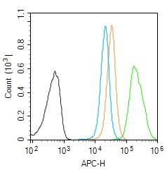
Fig2: Blank control (Black line): Molt4 (Black). Primary Antibody (green line):Rabbit Anti-DMT1 antibody (ER1912-90) Dilution: 1μg /10^6 cells; Isotype Control Antibody (orange line): Rabbit IgG . Secondary Antibody (white blue line): Goat anti-rabbit IgG-AF647 Dilution: 1μg /test. Protocol The cells were fixed with 4% PFA (10min at room temperature)and then permeabilized with PBST for 20 min at room temperature. The cells were then incubated in 5%BSA to block non-specific protein-protein interactions for 30 min at room temperature .Cells stained with Primary Antibody for 30 min at room temperature. The secondary antibody used for 40 min at room temperature. Acquisition of 20,000 events was performed.Application
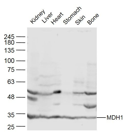
Fig3: Sample: Kidney (Mouse) Lysate at 40 ug Liver (Mouse) Lysate at 40 ug Heart (Mouse) Lysate at 40 ug Stomach (Mouse) Lysate at 40 ug Skin (Mouse) Lysate at 40 ug Bone (Mouse) Lysate at 40 ug Primary: Anti- MDH1 (ER1912-90) at 1/300 dilution Secondary: IRDye800CW Goat Anti-Rabbit IgG at 1/20000 dilution Predicted band size: 36 kD Observed band size: 36 kDApplication
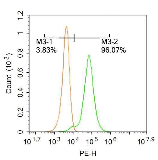
Fig4: Blank control:Molt-4. Primary Antibody (green line): Rabbit Anti-MDH1 antibody (ER1912-90) Dilution: 0.2μg /10^6 cells; Isotype Control Antibody (orange line): Rabbit IgG . Secondary Antibody : Goat anti-rabbit IgG-PE Dilution: 0.2μg /test. Protocol The cells were fixed with 4% PFA (10min at room temperature)and then permeabilized with 20% PBST for 20 min at-20℃. The cells were then incubated in 5%BSA to block non-specific protein-protein interactions for 30 min at at room temperature .Cells stained with Primary Antibody for 30 min at room temperature. The secondary antibody used for 40 min at room temperature. Acquisition of 20,000 events was performed.Application
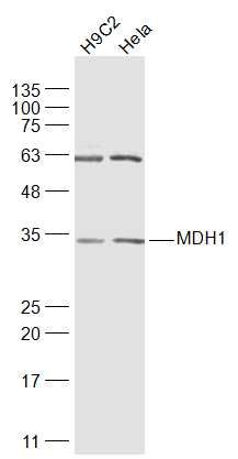
Fig5: Sample: H9C2(Rat) Cell Lysate at 30 ug Hela(Human) Cell Lysate at 30 ug Primary: Anti-MDH1 (ER1912-90) at 1/1000 dilution Secondary: IRDye800CW Goat Anti-Rabbit IgG at 1/20000 dilution Predicted band size: 36 kD Observed band size: 34 kDApplication
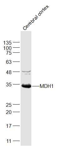
Fig6: Sample: Cerebral cortex(Rat) Lysate at 40 ug Primary: Anti-MDH1 (ER1912-90) at 1/1000 dilution Secondary: IRDye800CW Goat Anti-Rabbit IgG at 1/20000 dilution Predicted band size: 36 kD Observed band size: 34 kDApplication
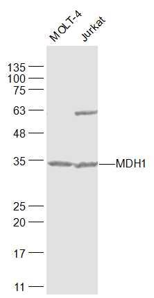
Fig7: Sample: MOLT-4(Human) Cell Lysate at 30 ug Jurkat(Human) Cell Lysate at 30 ug Primary: Anti-MDH1 (ER1912-90) at 1/1000 dilution Secondary: IRDye800CW Goat Anti-Rabbit IgG at 1/20000 dilution Predicted band size: 36 kD Observed band size: 34 kDApplication
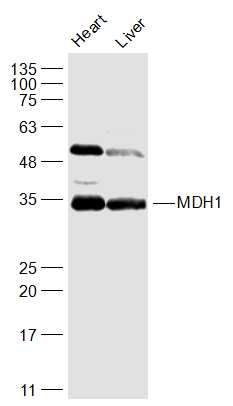
Fig8: Sample: Heart (Mouse) Lysate at 40 ug Liver (Mouse) Lysate at 40 ug Primary: Anti-MDH1 (ER1912-90) at 1/1000 dilution Secondary: IRDye800CW Goat Anti-Rabbit IgG at 1/20000 dilution Predicted band size: 36 kD Observed band size: 34 kD| Product Name | MDH1 |
|---|---|
| Antibody Type | Primary Antibodies |
| Antigen Alias | Cytosolic malate dehydrogenase Malate dehydrogenase 1, NAD (soluble) MDHA MDHs Soluble malate dehydrogenase Malate dehydrogenase Diiodophenylpyruvate reductase Malate dehydrogenase 1 Malate dehydrogenase 1 NAD (soluble) Malate dehydrogenase 2 Malate dehydrogenase cytoplasmic Malate dehydrogenase 2 NAD (mitochondrial) MDH 1 MDH 2 MDH MDH1 MDH2 MDHA MDHm MDHs MMDH Mor 1 Mor 2 Mor1 MOR2 cytoplasmic MDH s mdh1 MDHC_HUMAN MGC:1375 SMDH Soluble malate dehydrogenase. |
| Immunogen | KLH conjugated synthetic peptide derived from human MDH1 265-334/334 |
| Clonality | Polyclonal |
|---|---|
| Isotype | IgG |
| Host Species | Rabbit |
| Tested Applications | WBIHC-PFC |
| WB:1:500-2000: IHC-P:1:400-800: FC:0.2ug/Test: |
|
| Species Reactivity | HumanMouseRatChickenDogPigCowHorseRabbit |
| Concentration | 1mg/ml |
| Alternative Names | Cytosolic malate dehydrogenase
Malate dehydrogenase 1 NAD (soluble) MDHA MDHs Soluble malate dehydrogenase Malate dehydrogenase Diiodophenylpyruvate reductase Malate dehydrogenase 1 Malate dehydrogenase 1 NAD (soluble) Malate dehydrogenase 2 Malate dehydrogenase cytoplasmic Malate dehydrogenase 2 NAD (mitochondrial) MDH 1 MDH 2 MDH MDH1 MDH2 MDHA MDHm MDHs MMDH Mor 1 Mor 2 Mor1 MOR2 cytoplasmic MDH s mdh1 MDHC_HUMAN MGC:1375 SMDH Soluble malate dehydrogenase. |
|---|---|
| Molecular Weight(MW) | 36 KDa |
| Cellular Localization | Cytoplasm. |
| SwissProt ID | P40925 |
|---|
Application
Fig1: Paraformaldehyde-fixed, paraffin embedded (Rat brain); Antigen retrieval by microwave in sodium citrate buffer (pH6.0) ; Block endogenous peroxidase by 3% hydrogen peroxide for 30 minutes; Blocking buffer (3% BSA) at RT for 30min; Antibody incubation with (MDH1) Polyclonal Antibody, Unconjugated (ER1912-90) at 1:400 overnight at 4℃, followed by conjugation to the secondary antibody (labeled with HRP)and DAB staining.
Application
Fig2: Blank control (Black line): Molt4 (Black). Primary Antibody (green line):Rabbit Anti-DMT1 antibody (ER1912-90) Dilution: 1μg /10^6 cells; Isotype Control Antibody (orange line): Rabbit IgG . Secondary Antibody (white blue line): Goat anti-rabbit IgG-AF647 Dilution: 1μg /test. Protocol The cells were fixed with 4% PFA (10min at room temperature)and then permeabilized with PBST for 20 min at room temperature. The cells were then incubated in 5%BSA to block non-specific protein-protein interactions for 30 min at room temperature .Cells stained with Primary Antibody for 30 min at room temperature. The secondary antibody used for 40 min at room temperature. Acquisition of 20,000 events was performed.
Application
Fig3: Sample: Kidney (Mouse) Lysate at 40 ug Liver (Mouse) Lysate at 40 ug Heart (Mouse) Lysate at 40 ug Stomach (Mouse) Lysate at 40 ug Skin (Mouse) Lysate at 40 ug Bone (Mouse) Lysate at 40 ug Primary: Anti- MDH1 (ER1912-90) at 1/300 dilution Secondary: IRDye800CW Goat Anti-Rabbit IgG at 1/20000 dilution Predicted band size: 36 kD Observed band size: 36 kD
Application
Fig4: Blank control:Molt-4. Primary Antibody (green line): Rabbit Anti-MDH1 antibody (ER1912-90) Dilution: 0.2μg /10^6 cells; Isotype Control Antibody (orange line): Rabbit IgG . Secondary Antibody : Goat anti-rabbit IgG-PE Dilution: 0.2μg /test. Protocol The cells were fixed with 4% PFA (10min at room temperature)and then permeabilized with 20% PBST for 20 min at-20℃. The cells were then incubated in 5%BSA to block non-specific protein-protein interactions for 30 min at at room temperature .Cells stained with Primary Antibody for 30 min at room temperature. The secondary antibody used for 40 min at room temperature. Acquisition of 20,000 events was performed.
Application
Fig5: Sample: H9C2(Rat) Cell Lysate at 30 ug Hela(Human) Cell Lysate at 30 ug Primary: Anti-MDH1 (ER1912-90) at 1/1000 dilution Secondary: IRDye800CW Goat Anti-Rabbit IgG at 1/20000 dilution Predicted band size: 36 kD Observed band size: 34 kD
Application
Fig6: Sample: Cerebral cortex(Rat) Lysate at 40 ug Primary: Anti-MDH1 (ER1912-90) at 1/1000 dilution Secondary: IRDye800CW Goat Anti-Rabbit IgG at 1/20000 dilution Predicted band size: 36 kD Observed band size: 34 kD
Application
Fig7: Sample: MOLT-4(Human) Cell Lysate at 30 ug Jurkat(Human) Cell Lysate at 30 ug Primary: Anti-MDH1 (ER1912-90) at 1/1000 dilution Secondary: IRDye800CW Goat Anti-Rabbit IgG at 1/20000 dilution Predicted band size: 36 kD Observed band size: 34 kD
Application
Fig8: Sample: Heart (Mouse) Lysate at 40 ug Liver (Mouse) Lysate at 40 ug Primary: Anti-MDH1 (ER1912-90) at 1/1000 dilution Secondary: IRDye800CW Goat Anti-Rabbit IgG at 1/20000 dilution Predicted band size: 36 kD Observed band size: 34 kD| Application Notes | WB:1:500-2000: IHC-P:1:400-800: FC:0.2ug/Test: |
|---|
| Form | Liquid |
|---|---|
| Storage Instructions | Store at -20℃ for one year. Avoid repeated freeze/thaw cycles. The lyophilized antibody is stable at room temperature for at least one month and for greater than a year when kept at -20℃. When reconstituted in sterile pH 7.4 0.01M PBS or diluent of antibody the antibody is stable for at least two weeks at 2-4℃. |
| Storage Buffer | 0.01M TBS(pH7.4) with 1% BSA, 0.03% Proclin300 and 50% Glycerol. |


