Application
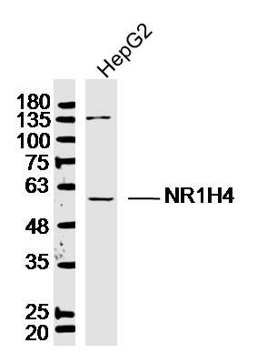
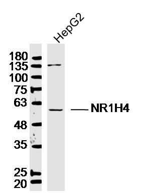
Fig1: Sample: HepG2 (Human) Cell Lysate at 30 ug Primary: Anti-NR1H4 (ER1914-12) at 1/300 dilution Secondary: IRDye800CW Goat Anti-Rabbit IgG at 1/20000 dilution Predicted band size: 56 kD Observed band size: 56 kDApplication
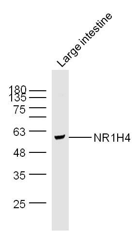
Fig2: Sample: Large intestine (Mouse) Lysate at 40 ug Primary: Anti-NR1H4 (ER1914-12) at 1/300 dilution Secondary: IRDye800CW Goat Anti-Rabbit IgG at 1/20000 dilution Predicted band size: 56 kD Observed band size: 56 kDApplication
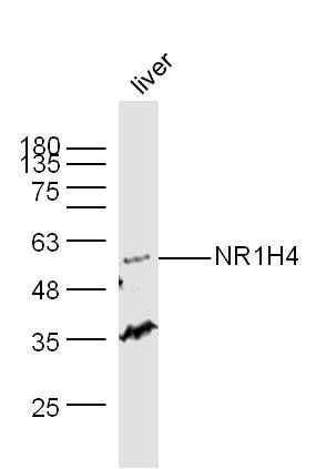
Fig3: Sample: Liver (Mouse) Lysate at 40 ug Primary: Anti-NR1H4 (ER1914-12) at 1/300 dilution Secondary: IRDye800CW Goat Anti-Rabbit IgG at 1/20000 dilution Predicted band size: 56 kD Observed band size: 56 kDApplication
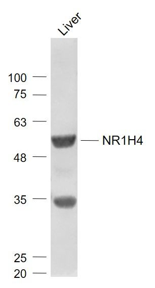
Fig4: Sample: Liver (Mouse) Lysate at 40 ug Primary: Anti- NR1H4 (ER1914-12) at 1/1000 dilution Secondary: IRDye800CW Goat Anti-Rabbit IgG at 1/20000 dilution Predicted band size: 56 kD Observed band size: 56 kDApplication
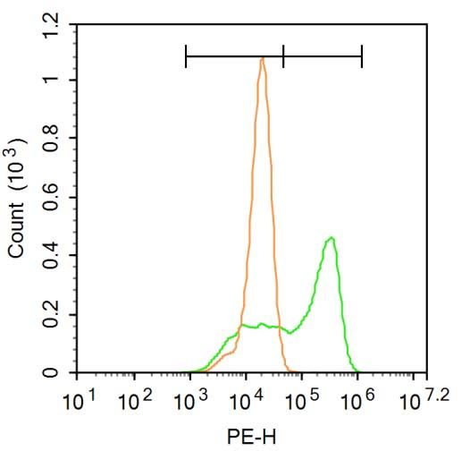
strong> Blank control:A549. Primary Antibody (green line): Rabbit Anti-NR1H4 antibody (ER1914-12) Dilution: 1μg /10^6 cells; Isotype Control Antibody (orange line): Rabbit IgG . Secondary Antibody : Goat anti-rabbit IgG-PE Dilution: 3μg /test. Protocol The cells were fixed with 4% PFA (10min at room temperature)and then permeabilized with 90% ice-cold methanol for 20 min at-20℃. The cells were then incubated in 5% BSA to block non-specific protein-protein interactions for 30 min at at room temperature .Cells stained with Primary Antibody for 30 min at room temperature. The secondary antibody used for 40 min at room temperature. Acquisition of 20,000 events was performed.Application
Fig6: Sample: HepG2 Cell (Human) Lysate at 40 ug Primary: Anti-NR1H4 (ER1914-12) at 1/300 dilution Secondary: IRDye800CW Goat Anti-Rabbit IgG at 1/20000 dilution Predicted band size: 56 kD Observed band size: 56 kD| Product Name | NR1H4 |
|---|---|
| Antibody Type | Primary Antibodies |
| Antigen Alias | Bile Acid Receptor NR1H4 BAR FXR Farnesoid X activated receptor Farnesoid X receptor Farnesoid X-activated receptor Farnesol receptor HRR 1 Farnesol receptor HRR-1 Farnesol receptor HRR1 FXR HRR 1 HRR1 NR1H4 NR1H4_HUMAN Nuclear receptor subfamily 1 group H member 4 Retinoid X receptor interacting protein 14 Retinoid X receptor-interacting protein 14 RIP 14 RIP14 RXR interacting protein 14 RXR-interacting protein 14. |
| Product description | This gene encodes a ligand-activated transcription factor that shares structural features in common with nuclear hormone receptor family members. This protein functions as a receptor for bile acids, and when bound to bile acids, binds to DNA and regulates the expression of genes involved in bile acid synthesis and transport. Alternatively spliced transcript variants encoding different isoforms have been described. Receptor for bile acids such as chenodeoxycholic acid, lithocholic acid and deoxycholic acid. Represses the transcription of the cholesterol 7-alpha-hydroxylase gene (CYP7A1) through the induction of NR0B2 or FGF19 expression, via two distinct mechanisms. Activates the intestinal bile acid-binding protein (IBABP). Activates the transcription of bile salt export pump ABCB11 by directly recruiting histone methyltransferase CARM1 to this locus. |
| Immunogen | KLH conjugated synthetic peptide derived from human FXR/Bile Acid Receptor NR1H4 175-280/486 |
| Clonality | Polyclonal |
|---|---|
| Isotype | IgG |
| Host Species | Rabbit |
| Tested Applications | WBFC |
| WB:1:500-2000: FC:3ug/test: |
|
| Species Reactivity | HumanMouseRatDogPigCowHorseSheep |
| Concentration | 1mg/ml |
| Alternative Names | Bile Acid Receptor NR1H4
BAR
FXR Farnesoid X activated receptor Farnesoid X receptor Farnesoid X-activated receptor Farnesol receptor HRR 1 Farnesol receptor HRR-1 Farnesol receptor HRR1 FXR HRR 1 HRR1 NR1H4 NR1H4_HUMAN Nuclear receptor subfamily 1 group H member 4 Retinoid X receptor interacting protein 14 Retinoid X receptor-interacting protein 14 RIP 14 RIP14 RXR interacting protein 14 RXR-interacting protein 14. |
|---|---|
| Molecular Weight(MW) | 56 KDa |
| Cellular Localization | Nucleus. |
| SwissProt ID | Q96RI1 |
|---|
Application
Fig1: Sample: HepG2 (Human) Cell Lysate at 30 ug Primary: Anti-NR1H4 (ER1914-12) at 1/300 dilution Secondary: IRDye800CW Goat Anti-Rabbit IgG at 1/20000 dilution Predicted band size: 56 kD Observed band size: 56 kD
Application
Fig2: Sample: Large intestine (Mouse) Lysate at 40 ug Primary: Anti-NR1H4 (ER1914-12) at 1/300 dilution Secondary: IRDye800CW Goat Anti-Rabbit IgG at 1/20000 dilution Predicted band size: 56 kD Observed band size: 56 kD
Application
Fig3: Sample: Liver (Mouse) Lysate at 40 ug Primary: Anti-NR1H4 (ER1914-12) at 1/300 dilution Secondary: IRDye800CW Goat Anti-Rabbit IgG at 1/20000 dilution Predicted band size: 56 kD Observed band size: 56 kD
Application
Fig4: Sample: Liver (Mouse) Lysate at 40 ug Primary: Anti- NR1H4 (ER1914-12) at 1/1000 dilution Secondary: IRDye800CW Goat Anti-Rabbit IgG at 1/20000 dilution Predicted band size: 56 kD Observed band size: 56 kD
Application
strong> Blank control:A549. Primary Antibody (green line): Rabbit Anti-NR1H4 antibody (ER1914-12) Dilution: 1μg /10^6 cells; Isotype Control Antibody (orange line): Rabbit IgG . Secondary Antibody : Goat anti-rabbit IgG-PE Dilution: 3μg /test. Protocol The cells were fixed with 4% PFA (10min at room temperature)and then permeabilized with 90% ice-cold methanol for 20 min at-20℃. The cells were then incubated in 5% BSA to block non-specific protein-protein interactions for 30 min at at room temperature .Cells stained with Primary Antibody for 30 min at room temperature. The secondary antibody used for 40 min at room temperature. Acquisition of 20,000 events was performed.
Application
Fig6: Sample: HepG2 Cell (Human) Lysate at 40 ug Primary: Anti-NR1H4 (ER1914-12) at 1/300 dilution Secondary: IRDye800CW Goat Anti-Rabbit IgG at 1/20000 dilution Predicted band size: 56 kD Observed band size: 56 kD| Positive Control | HepG2 Cell Lysate, Mouse Large intestine Lysate, Mouse Liver Lysate, Mouse Liver Lysate, A549 cell, HepG2 Cell Lysate. |
|---|---|
| Application Notes | WB:1:500-2000: FC:3ug/test: |
| Form | Liquid |
|---|---|
| Storage Instructions | Store at -20℃ for one year. Avoid repeated freeze/thaw cycles. The lyophilized antibody is stable at room temperature for at least one month and for greater than a year when kept at -20℃. When reconstituted in sterile pH 7.4 0.01M PBS or diluent of antibody the antibody is stable for at least two weeks at 2-4℃. |
| Storage Buffer | 0.01M TBS(pH7.4) with 1% BSA, 0.03% Proclin300 and 50% Glycerol. |


