Application
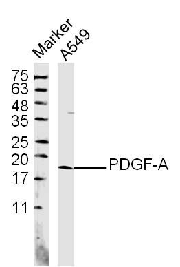
Fig1: Sample: A549 (human)Cell Lysate at 40 ug Primary: Anti- PDGF-A (ER1914-91) at 1/300 dilution Secondary: IRDye800CW Goat Anti-Rabbit IgG at 1/20000 dilution Predicted band size: 14 kD Observed band size: 18 kDApplication
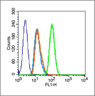
Fig2: Blank control (blue line): A431 cells (blue). Primary Antibody (green line): Rabbit Anti-PDGF-A antibody (ER1914-91) Dilution: 1μg /10^6 cells; Isotype Control Antibody (orange line): Rabbit IgG . Secondary Antibody (white blue line): Goat anti-rabbit IgG-FITC Dilution: 1μg /test. Protocol The cells were fixed with 70% methanol (Overnight at 4℃) and then permeabilized with 90% ice-cold methanol for 20 min at -20℃. Cells stained with Primary Antibody for 30 min at room temperature. The cells were then incubated in 1 X PBS/2%BSA/10% goat serum to block non-specific protein-protein interactions followed by the antibody for 15 min at room temperature. The secondary antibody used for 40 min at room temperature. Acquisition of 20,000 events was performed.Application
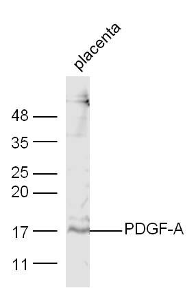
Fig3: Sample: placenta (Mouse) Lysate at 40 ug Primary: Anti-PDGF-A (ER1914-91) at 1/300 dilution Secondary: IRDye800CW Goat Anti-Rabbit IgG at 1/20000 dilution Predicted band size: 14 kD Observed band size: 17 kD| Product Name | PDGF-A |
|---|---|
| Antibody Type | Primary Antibodies |
| Antigen Alias | Platelet–dirived growth factor–1 PDGFA PDGF AA PDGF-AA PDGF A chain PDGF subunit A PDGF-1 PDGF1 PDGFA PDGFA_HUMAN Platelet derived growth factor A chain precursor Platelet derived growth factor alpha polypeptide Platelet-derived growth factor A chain Platelet-derived growth factor alpha polypeptide Platelet-derived growth factor subunit A. |
| Product description | Growth factor that plays an essential role in the regulation of embryonic development, cell proliferation, cell migration, survival and chemotaxis. Potent mitogen for cells of mesenchymal origin. Required for normal lung alveolar septum formation during embryogenesis, normal development of the gastrointestinal tract, normal development of Leydig cells and spermatogenesis. Required for normal oligodendrocyte development and normal myelination in the spinal cord and cerebellum. Plays an important role in wound healing. Signaling is modulated by the formation of heterodimers with PDGFB. |
| Immunogen | KLH conjugated synthetic peptide derived from the missle of human PDGF-A 125-170/211 |
| Clonality | Polyclonal |
|---|---|
| Isotype | IgG |
| Host Species | Rabbit |
| Tested Applications | WBFC |
| WB:1:500-2000: FC:1μg/Test: |
|
| Species Reactivity | HumanMouseRatRabbit |
| Concentration | 1mg/ml |
| Alternative Names | Platelet–dirived growth factor–1 PDGFA PDGF AA PDGF-AA PDGF A chain PDGF subunit A PDGF-1 PDGF1 PDGFA PDGFA_HUMAN Platelet derived growth factor A chain precursor Platelet derived growth factor alpha polypeptide Platelet-derived growth factor A chain Platelet-derived growth factor alpha polypeptide Platelet-derived growth factor subunit A. |
|---|---|
| Molecular Weight(MW) | 14 KDa |
| Cellular Localization | Secreted. Note=Released by platelets upon wounding. |
| SwissProt ID | P04085 |
|---|
Application
Fig1: Sample: A549 (human)Cell Lysate at 40 ug Primary: Anti- PDGF-A (ER1914-91) at 1/300 dilution Secondary: IRDye800CW Goat Anti-Rabbit IgG at 1/20000 dilution Predicted band size: 14 kD Observed band size: 18 kD
Application
Fig2: Blank control (blue line): A431 cells (blue). Primary Antibody (green line): Rabbit Anti-PDGF-A antibody (ER1914-91) Dilution: 1μg /10^6 cells; Isotype Control Antibody (orange line): Rabbit IgG . Secondary Antibody (white blue line): Goat anti-rabbit IgG-FITC Dilution: 1μg /test. Protocol The cells were fixed with 70% methanol (Overnight at 4℃) and then permeabilized with 90% ice-cold methanol for 20 min at -20℃. Cells stained with Primary Antibody for 30 min at room temperature. The cells were then incubated in 1 X PBS/2%BSA/10% goat serum to block non-specific protein-protein interactions followed by the antibody for 15 min at room temperature. The secondary antibody used for 40 min at room temperature. Acquisition of 20,000 events was performed.
Application
Fig3: Sample: placenta (Mouse) Lysate at 40 ug Primary: Anti-PDGF-A (ER1914-91) at 1/300 dilution Secondary: IRDye800CW Goat Anti-Rabbit IgG at 1/20000 dilution Predicted band size: 14 kD Observed band size: 17 kD| Positive Control | Platelet-derived growth factor is a potent mitogen for cells of mesenchymal origin. Binding of this growth factor to its receptor elicits a variety of cellular responses. It is released by platelets upon wounding and plays an important role in stimulating adjacent cells to grow and thereby heals the wound. |
|---|---|
| Application Notes | WB:1:500-2000: FC:1μg/Test: |
| Form | Liquid |
|---|---|
| Storage Instructions | Store at -20℃ for one year. Avoid repeated freeze/thaw cycles. The lyophilized antibody is stable at room temperature for at least one month and for greater than a year when kept at -20℃. When reconstituted in sterile pH 7.4 0.01M PBS or diluent of antibody the antibody is stable for at least two weeks at 2-4℃. |
| Storage Buffer | 0.01M TBS(pH7.4) with 1% BSA, 0.03% Proclin300 and 50% Glycerol. |


