Application
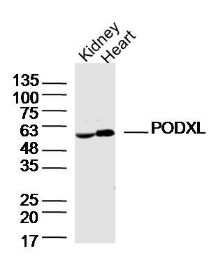
Fig1: Sample: Kidney(Mouse)Lysate at 40 ug Heart (Mouse)Lysate at 40 ug Primary: Anti-PODXL(ER1915-24)at 1/300 dilution Secondary: IRDye800CW Goat Anti-RabbitIgG at 1/20000 dilution Predicted band size: 59kD Observed band size: 59kD| Product Name | PODXL |
|---|---|
| Antibody Type | Primary Antibodies |
| Antigen Alias | Podocalyxin PCLP PCLP1 podocalyxin-like isoform 1 precursor Podocalyxin like protein PODXL_MOUSE GCTM-2 antigen Gp200 Podocalyxin-like protein 1 PC PCLP-1 PCX PODXL. |
| Product description | Involved in the regulation of both adhesion and cell morphology and cancer progression. Function as an anti-adhesive molecule that maintains an open filtration pathway between neighboring foot processes in the podocyte by charge repulsion. Acts as a pro-adhesive molecule, enhancing the adherence of cells to immobilized ligands, increasing the rate of migration and cell-cell contacts in an integrin-dependent manner. Induces the formation of apical actin-dependent microvilli. Involved in the formation of a preapical plasma membrane subdomain to set up inital epithelial polarization and the apical lumen formation during renal tubulogenesis. Plays a role in cancer development and aggressiveness by inducing cell migration and invasion through its interaction with the actin-binding protein EZR. Affects EZR-dependent signaling events, leading to increased activities of the MAPK and PI3K pathways in cancer cells. |
| Immunogen | KLH conjugated synthetic peptide derived from mouse PODXL 351-450/558 |
| Clonality | Polyclonal |
|---|---|
| Isotype | IgG |
| Host Species | Rabbit |
| Tested Applications | WB |
| WB:1:500-2000: |
|
| Species Reactivity | MouseRat |
| Concentration | 1mg/ml |
| Alternative Names | Podocalyxin PCLP PCLP1 podocalyxin-like isoform 1 precursor Podocalyxin like protein PODXL_MOUSE GCTM-2 antigen Gp200 Podocalyxin-like protein 1 PC PCLP-1 PCX PODXL. |
|---|---|
| Molecular Weight(MW) | 59 KDa |
| Cellular Localization | Apical cell membrane. Cell projection, lamellipodium. Cell projection, filopodium. Cell projection, ruffle. Cell projection, microvillus. Membrane raft. Membrane. In single attached epithelial cells is restricted to a preapical pole on the free plasma membrane whereas other apical and basolateral proteins are not yet polarized. Colocalizes with SLC9A3R2 at the apical plasma membrane during epithelial polarization. Colocalizes with SLC9A3R1 at the trans-Golgi network (transiently) and at the apical plasma membrane. Its association with the membrane raft is transient. Colocalizes with actin filaments, EZR and SLC9A3R1 in a punctate pattern at the apical cell surface where microvilli form. Colocalizes with EZR and SLC9A3R2 at the apical cell membrane of glomerular epithelium cells (By similarity). Forms granular, punctuated pattern, forming patches, preferentially adopting a polar distribution, located on the migrating poles of the cell or forming clusters along the terminal ends of filipodia establishing contact with the endothelial cells. Colocalizes with the submembrane actin of lamellipodia, particularly associated with ruffles. Colocalizes with vinculin at protrusions of cells. Colocalizes with ITGB1. Colocalizes with PARD3, PRKCI, EXOC5, OCLN, RAB11A and RAB8A in apical membrane initiation sites (AMIS) during the generation of apical surface and luminogenesis. |
| SwissProt ID | Q9R0M4 |
|---|
Application
Fig1: Sample: Kidney(Mouse)Lysate at 40 ug Heart (Mouse)Lysate at 40 ug Primary: Anti-PODXL(ER1915-24)at 1/300 dilution Secondary: IRDye800CW Goat Anti-RabbitIgG at 1/20000 dilution Predicted band size: 59kD Observed band size: 59kD| Positive Control | Glomerular epithelium cell (podocyte). |
|---|---|
| Application Notes | WB:1:500-2000: |
| Form | Liquid |
|---|---|
| Storage Instructions | Store at -20℃ for one year. Avoid repeated freeze/thaw cycles. The lyophilized antibody is stable at room temperature for at least one month and for greater than a year when kept at -20℃. When reconstituted in sterile pH 7.4 0.01M PBS or diluent of antibody the antibody is stable for at least two weeks at 2-4℃. |
| Storage Buffer | 0.01M TBS(pH7.4) with 1% BSA, 0.03% Proclin300 and 50% Glycerol. |


