Application
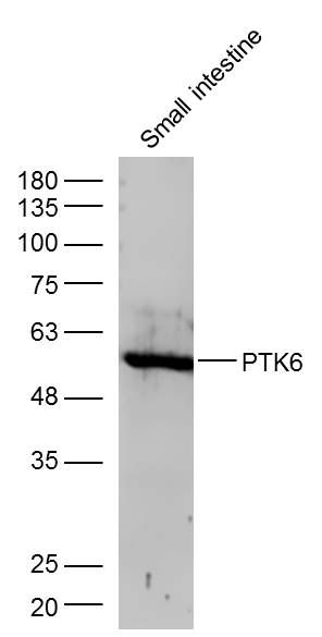
Fig1: Sample: Small intestine (Mouse) Lysate at 40 ug Primary: Anti- PTK6 (ER1915-59) at 1/300 dilution Secondary: IRDye800CW Goat Anti-Rabbit IgG at 1/20000 dilution Predicted band size: 52 kD Observed band size: 52 kD| Product Name | PTK6 |
|---|---|
| Antibody Type | Primary Antibodies |
| Antigen Alias | Breast tumor kinase Breast tumour kinase Protein tyrosine kinase 6 PTK6 Tyrosine protein kinase BRK PTK6_HUMAN. |
| Product description | Non-receptor tyrosine-protein kinase implicated in the regulation of a variety of signaling pathways that control the differentiation and maintenance of normal epithelia, as well as tumor growth. Function seems to be context dependent and differ depending on cell type, as well as its intracellular localization. A number of potential nuclear and cytoplasmic substrates have been identified. These include the RNA-binding proteins: KHDRBS1/SAM68, KHDRBS2/SLM1, KHDRBS3/SLM2 and SFPQ/PSF; transcription factors: STAT3 and STAT5A/B and a variety of signaling molecules: ARHGAP35/p190RhoGAP, PXN/paxillin, BTK/ATK, STAP2/BKS. Associates also with a variety of proteins that are likely upstream of PTK6 in various signaling pathways, or for which PTK6 may play an adapter-like role. These proteins include ADAM15, EGFR, ERBB2, ERBB3 and IRS4. In normal or non-tumorigenic tissues, PTK6 promotes cellular differentiation and apoptosis. In tumors PTK6 contributes to cancer progression by sensitizing cells to mitogenic signals and enhancing proliferation, anchorage-independent survival and migration/invasion. Association with EGFR, ERBB2, ERBB3 may contribute to mammary tumor development and growth through enhancement of EGF-induced signaling via BTK/AKT and PI3 kinase. Contributes to migration and proliferation by contributing to EGF-mediated phosphorylation of ARHGAP35/p190RhoGAP, which promotes association with RASA1/p120RasGAP, inactivating RhoA while activating RAS. EGF stimulation resulted in phosphorylation of PNX/Paxillin by PTK6 and activation of RAC1 via CRK/CrKII, thereby promoting migration and invasion. PTK6 activates STAT3 and STAT5B to promote proliferation. Nuclear PTK6 may be important for regulating growth in normal epithelia, while cytoplasmic PTK6 might activate oncogenic signaling pathways. ;Isoform 2 inhibits PTK6 phosphorylation and PTK6 association with other tyrosine-phosphorylated proteins. |
| Immunogen | KLH conjugated synthetic peptide derived from human Brk/PTK6 201-300/451 |
| Clonality | Polyclonal |
|---|---|
| Isotype | IgG |
| Host Species | Rabbit |
| Tested Applications | WB |
| WB:1:500-2000: |
|
| Species Reactivity | HumanMouseRatCowSheep |
| Concentration | 1mg/ml |
| Alternative Names | Breast tumor kinase Breast tumour kinase Protein tyrosine kinase 6 PTK6 Tyrosine protein kinase BRK PTK6_HUMAN. |
|---|---|
| Molecular Weight(MW) | 52 KDa |
| Cellular Localization | Cytoplasm. Nucleus. Cell projection, ruffle. Membrane (By similarity). Note=Colocalizes with KHDRBS1, KHDRBS2 or KHDRBS3, within the nucleus. Nuclear localization in epithelial cells of normal prostate but cytoplasmic localization in cancer prostate. |
| SwissProt ID | Q13882 |
|---|
Application
Fig1: Sample: Small intestine (Mouse) Lysate at 40 ug Primary: Anti- PTK6 (ER1915-59) at 1/300 dilution Secondary: IRDye800CW Goat Anti-Rabbit IgG at 1/20000 dilution Predicted band size: 52 kD Observed band size: 52 kD| Positive Control | Epithelia-specific. Very high level in colon and high levels in small intestine and prostate, and low levels in some fetal tissues. Not expressed in breast or ovarian tissue but expressed in high pourcentage of breast and ovarian cancers. Also overexpressed in some metastatic melanomas, lymphomas, colon cancers, squamous cell carcinomas and prostate cancers. Also found in melanocytes. Not expressed in heart, brain, placenta, lung, liver, skeletal muscle, kidney and pancreas. Isoform 2 is present in prostate epithelial cell lines derived from normal prostate and prostate adenocarcinomas, as well as in a variety of cell lines. |
|---|---|
| Application Notes | WB:1:500-2000: |
| Form | Liquid |
|---|---|
| Storage Instructions | Store at -20℃ for one year. Avoid repeated freeze/thaw cycles. The lyophilized antibody is stable at room temperature for at least one month and for greater than a year when kept at -20℃. When reconstituted in sterile pH 7.4 0.01M PBS or diluent of antibody the antibody is stable for at least two weeks at 2-4℃. |
| Storage Buffer | 0.01M TBS(pH7.4) with 1% BSA, 0.03% Proclin300 and 50% Glycerol. |


