Application
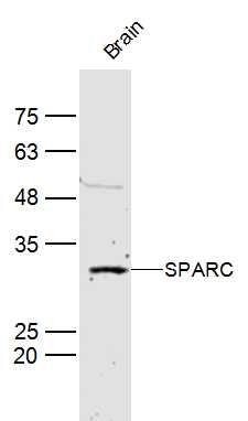
Fig1: Sample: Brain (Mouse) Lysate at 40 ug Primary: Anti-SPARC (ER1916-99) at 1/300 dilution Secondary: IRDye800CW Goat Anti-Rabbit IgG at 1/20000 dilution Predicted band size: 33 kD Observed band size: 33 kDApplication
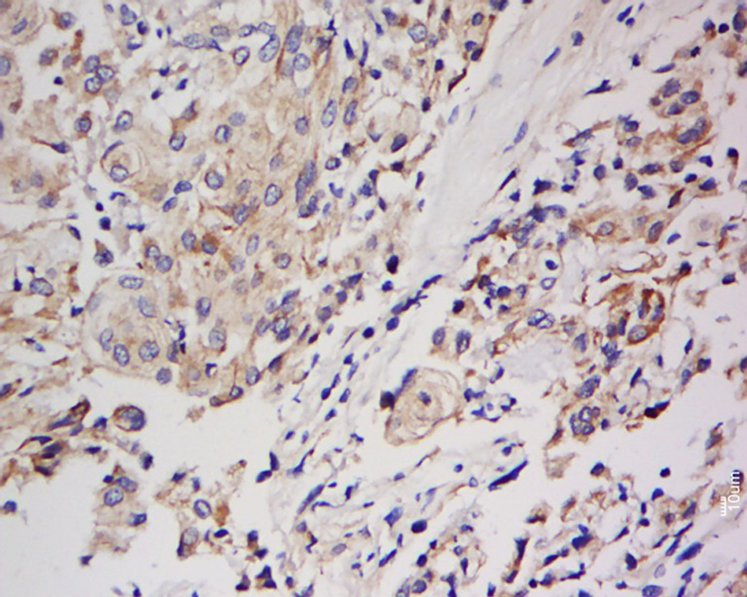
Fig2: Tissue/cell: Human Meningioma; 4% Paraformaldehyde-fixed and paraffin-embedded; Antigen retrieval: citrate buffer ( 0.01M, pH 6.0 ), Boiling bathing for 15min; Block endogenous peroxidase by 3% Hydrogen peroxide for 30min; Blocking buffer (normal goat serum,C-0005) at 37℃ for 20 min; Incubation: Anti-SPARC Polyclonal Antibody, Unconjugated(ER1916-99) 1:200, overnight at 4℃, followed by conjugation to the secondary antibody(SP-0023) and DAB(C-0010) stainingApplication
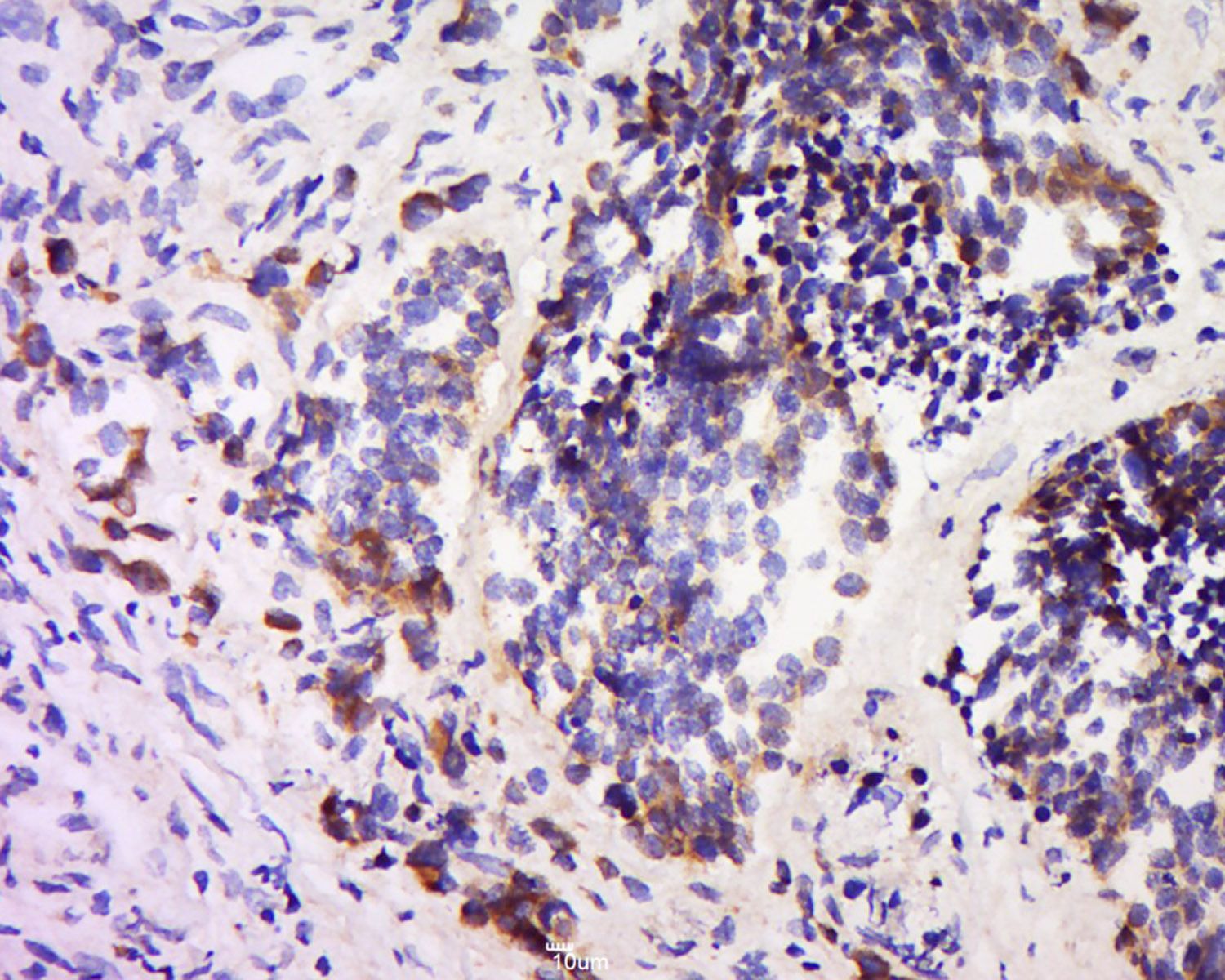
Fig3: Tissue/cell: Human colon cancer; 4% Paraformaldehyde-fixed and paraffin-embedded; Antigen retrieval: citrate buffer ( 0.01M, pH 6.0 ), Boiling bathing for 15min; Block endogenous peroxidase by 3% Hydrogen peroxide for 30min; Blocking buffer (normal goat serum,C-0005) at 37℃ for 20 min; Incubation: Anti-SPARC Polyclonal Antibody, Unconjugated(ER1916-99) 1:200, overnight at 4℃, followed by conjugation to the secondary antibody(SP-0023) and DAB(C-0010) stainingApplication
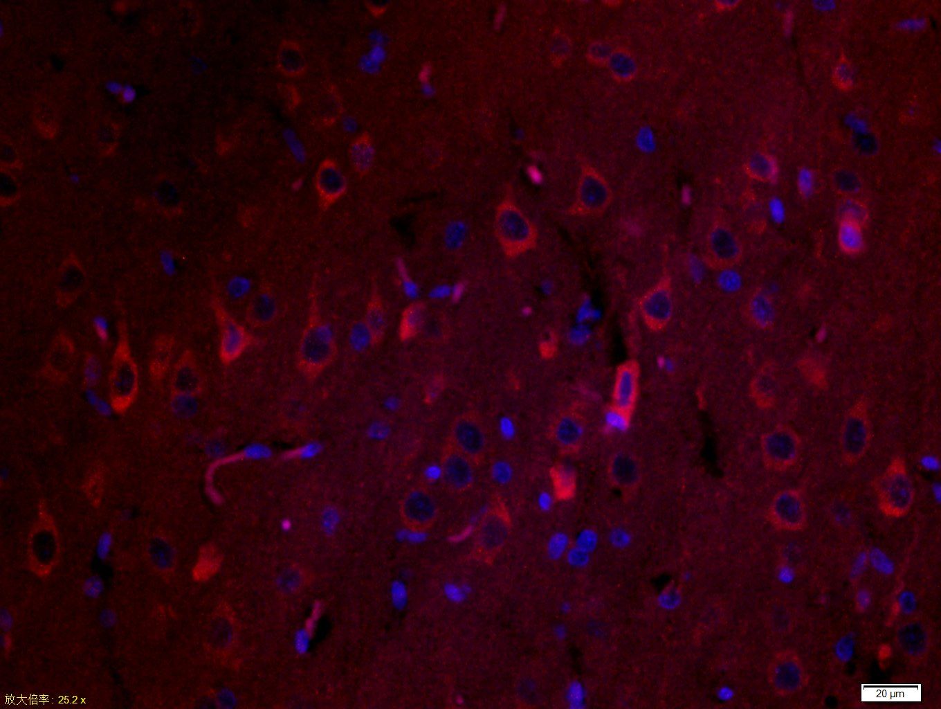
Fig4: Tissue/cell: rat brain tissue;4% Paraformaldehyde-fixed and paraffin-embedded; Antigen retrieval: citrate buffer ( 0.01M, pH 6.0 ), Boiling bathing for 15min; Blocking buffer (normal goat serum,C-0005) at 37℃ for 20 min; Incubation: Anti-SPARC Polyclonal Antibody, Unconjugated(ER1916-99) 1:200, overnight at 4℃; The secondary antibody was Goat Anti-Rabbit IgG, Cy3 conjugated(bs-0295G-Cy3)used at 1:200 dilution for 40 minutes at 37℃. DAPI(5ug/ml,blue,C-0033) was used to stain the cell nucleiApplication
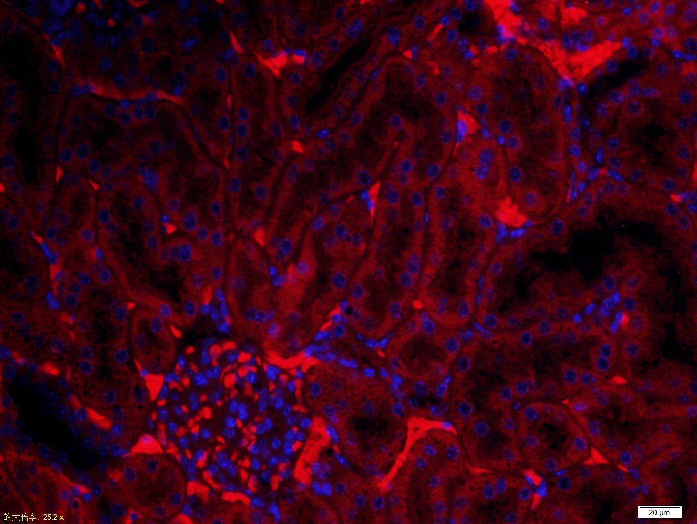
Fig5: Tissue/cell: mouse kidney tissue;4% Paraformaldehyde-fixed and paraffin-embedded; Antigen retrieval: citrate buffer ( 0.01M, pH 6.0 ), Boiling bathing for 15min; Blocking buffer (normal goat serum,C-0005) at 37℃ for 20 min; Incubation: Anti-SPARC Polyclonal Antibody, Unconjugated(ER1916-99) 1:200, overnight at 4℃; The secondary antibody was Goat Anti-Rabbit IgG, Cy3 conjugated(bs-0295G-Cy3)used at 1:200 dilution for 40 minutes at 37℃. DAPI(5ug/ml,blue,C-0033) was used to stain the cell nucleiApplication
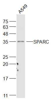
Fig6: Sample: A549(Human) Cell Lysate at 30 ug Primary: Anti-SPARC (ER1916-99) at 1/1000 dilution Secondary: IRDye800CW Goat Anti-Rabbit IgG at 1/20000 dilution Predicted band size: 33 kD Observed band size: 35 kDApplication
Fig7: Paraformaldehyde-fixed, paraffin embedded (human brain glioma); Antigen retrieval by boiling in sodium citrate buffer (pH6.0) for 15min; Block endogenous peroxidase by 3% hydrogen peroxide for 20 minutes; Blocking buffer (normal goat serum) at 37℃ for 30min; Antibody incubation with (SPARC) Polyclonal Antibody, Unconjugated (ER1916-99) at 1:400 overnight at 4℃, followed by operating according to SP Kit(Rabbit) (sp-0023) instructionsand DAB staining.Application
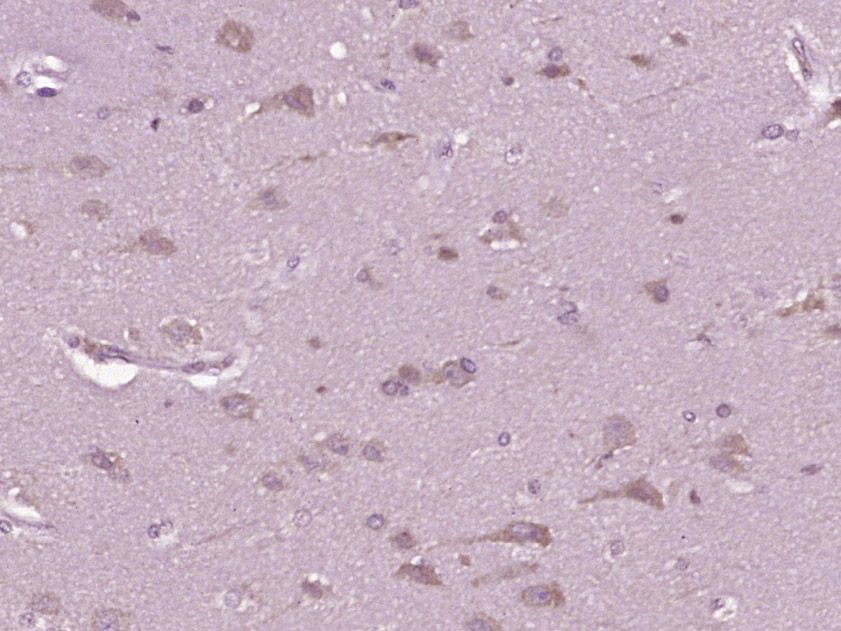
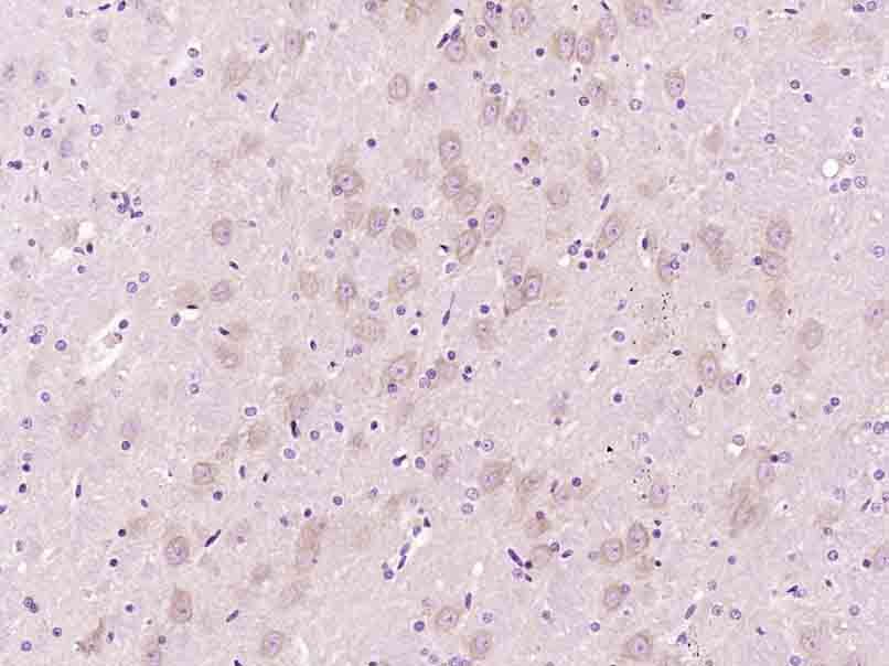
Fig8: Paraformaldehyde-fixed, paraffin embedded (mouse brain tissue); Antigen retrieval by boiling in sodium citrate buffer (pH6.0) for 15min; Block endogenous peroxidase by 3% hydrogen peroxide for 20 minutes; Blocking buffer (normal goat serum) at 37℃ for 30min; Antibody incubation with (SPARC) Polyclonal Antibody, Unconjugated (ER1916-99) at 1:400 overnight at 4℃, followed by operating according to SP Kit(Rabbit) (sp-0023) instructionsand DAB staining.Application
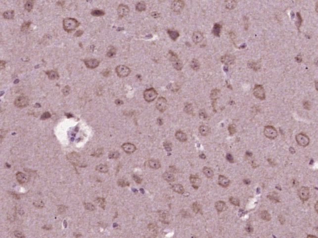
Fig9: Paraformaldehyde-fixed, paraffin embedded (Rat brain); Antigen retrieval by boiling in sodium citrate buffer (pH6.0) for 15min; Block endogenous peroxidase by 3% hydrogen peroxide for 20 minutes; Blocking buffer (normal goat serum) at 37℃ for 30min; Antibody incubation with (SPARC) Polyclonal Antibody, Unconjugated (ER1916-99) at 1:400 overnight at 4℃, followed by operating according to SP Kit(Rabbit) (sp-0023) instructionsand DAB staining.| Product Name | SPARC |
|---|---|
| Antibody Type | Primary Antibodies |
| Antigen Alias | AA517111 Basement membrane protein 40 BM 40 BM40 Cysteine rich protein hm:zeh0062 MGC128090 ON Osteonectin Secreted acidic cystein rich glycoprotein Secreted protein acidic and rich in cysteine Secreted protein acidic cysteine rich (osteonectin) Secreted protein acidic cysteine rich SPRC SPRC_HUMAN. |
| Product description | This gene encodes a cysteine-rich acidic matrix-associated protein. The encoded protein is required for the collagen in bone to become calcified but is also involved in extracellular matrix synthesis and promotion of changes to cell shape. The gene product has been associated with tumor suppression but has also been correlated with metastasis based on changes to cell shape which can promote tumor cell invasion. Three transcript variants encoding different isoforms have been found for this gene. Appears to regulate cell growth through interactions with the extracellular matrix and cytokines. Binds calcium and copper, several types of collagen, albumin, thrombospondin, PDGF and cell membranes. There are two calcium binding sites; an acidic domain that binds 5 to 8 Ca(2+) with a low affinity and an EF-hand loop that binds a Ca(2+) ion with a high affinity. |
| Immunogen | KLH conjugated synthetic peptide derived from human SPARC 101-200/303 |
| Clonality | Polyclonal |
|---|---|
| Isotype | IgG |
| Host Species | Rabbit |
| Tested Applications | WBIHC-PICC/IF |
| WB:1:500-2000: IHC-P:1:400-800: IF:1:100-500: |
|
| Species Reactivity | HumanMouseRatDogPigCowHorseRabbit |
| Concentration | 1mg/ml |
| Alternative Names | AA517111
Basement membrane protein 40
BM 40 BM40 Cysteine rich protein hm:zeh0062 MGC128090 ON Osteonectin Secreted acidic cystein rich glycoprotein Secreted protein acidic and rich in cysteine Secreted protein acidic cysteine rich (osteonectin) Secreted protein acidic cysteine rich SPRC SPRC_HUMAN. |
|---|---|
| Molecular Weight(MW) | 33 KDa |
| Cellular Localization | Secreted, extracellular space, extracellular matrix, basement membrane. Note=In or around the basement membrane. |
| SwissProt ID | P09486 |
|---|
Application
Fig1: Sample: Brain (Mouse) Lysate at 40 ug Primary: Anti-SPARC (ER1916-99) at 1/300 dilution Secondary: IRDye800CW Goat Anti-Rabbit IgG at 1/20000 dilution Predicted band size: 33 kD Observed band size: 33 kD
Application
Fig2: Tissue/cell: Human Meningioma; 4% Paraformaldehyde-fixed and paraffin-embedded; Antigen retrieval: citrate buffer ( 0.01M, pH 6.0 ), Boiling bathing for 15min; Block endogenous peroxidase by 3% Hydrogen peroxide for 30min; Blocking buffer (normal goat serum,C-0005) at 37℃ for 20 min; Incubation: Anti-SPARC Polyclonal Antibody, Unconjugated(ER1916-99) 1:200, overnight at 4℃, followed by conjugation to the secondary antibody(SP-0023) and DAB(C-0010) staining
Application
Fig3: Tissue/cell: Human colon cancer; 4% Paraformaldehyde-fixed and paraffin-embedded; Antigen retrieval: citrate buffer ( 0.01M, pH 6.0 ), Boiling bathing for 15min; Block endogenous peroxidase by 3% Hydrogen peroxide for 30min; Blocking buffer (normal goat serum,C-0005) at 37℃ for 20 min; Incubation: Anti-SPARC Polyclonal Antibody, Unconjugated(ER1916-99) 1:200, overnight at 4℃, followed by conjugation to the secondary antibody(SP-0023) and DAB(C-0010) staining
Application
Fig4: Tissue/cell: rat brain tissue;4% Paraformaldehyde-fixed and paraffin-embedded; Antigen retrieval: citrate buffer ( 0.01M, pH 6.0 ), Boiling bathing for 15min; Blocking buffer (normal goat serum,C-0005) at 37℃ for 20 min; Incubation: Anti-SPARC Polyclonal Antibody, Unconjugated(ER1916-99) 1:200, overnight at 4℃; The secondary antibody was Goat Anti-Rabbit IgG, Cy3 conjugated(bs-0295G-Cy3)used at 1:200 dilution for 40 minutes at 37℃. DAPI(5ug/ml,blue,C-0033) was used to stain the cell nuclei
Application
Fig5: Tissue/cell: mouse kidney tissue;4% Paraformaldehyde-fixed and paraffin-embedded; Antigen retrieval: citrate buffer ( 0.01M, pH 6.0 ), Boiling bathing for 15min; Blocking buffer (normal goat serum,C-0005) at 37℃ for 20 min; Incubation: Anti-SPARC Polyclonal Antibody, Unconjugated(ER1916-99) 1:200, overnight at 4℃; The secondary antibody was Goat Anti-Rabbit IgG, Cy3 conjugated(bs-0295G-Cy3)used at 1:200 dilution for 40 minutes at 37℃. DAPI(5ug/ml,blue,C-0033) was used to stain the cell nuclei
Application
Fig6: Sample: A549(Human) Cell Lysate at 30 ug Primary: Anti-SPARC (ER1916-99) at 1/1000 dilution Secondary: IRDye800CW Goat Anti-Rabbit IgG at 1/20000 dilution Predicted band size: 33 kD Observed band size: 35 kD
Application
Fig7: Paraformaldehyde-fixed, paraffin embedded (human brain glioma); Antigen retrieval by boiling in sodium citrate buffer (pH6.0) for 15min; Block endogenous peroxidase by 3% hydrogen peroxide for 20 minutes; Blocking buffer (normal goat serum) at 37℃ for 30min; Antibody incubation with (SPARC) Polyclonal Antibody, Unconjugated (ER1916-99) at 1:400 overnight at 4℃, followed by operating according to SP Kit(Rabbit) (sp-0023) instructionsand DAB staining.
Application
Fig8: Paraformaldehyde-fixed, paraffin embedded (mouse brain tissue); Antigen retrieval by boiling in sodium citrate buffer (pH6.0) for 15min; Block endogenous peroxidase by 3% hydrogen peroxide for 20 minutes; Blocking buffer (normal goat serum) at 37℃ for 30min; Antibody incubation with (SPARC) Polyclonal Antibody, Unconjugated (ER1916-99) at 1:400 overnight at 4℃, followed by operating according to SP Kit(Rabbit) (sp-0023) instructionsand DAB staining.
Application
Fig9: Paraformaldehyde-fixed, paraffin embedded (Rat brain); Antigen retrieval by boiling in sodium citrate buffer (pH6.0) for 15min; Block endogenous peroxidase by 3% hydrogen peroxide for 20 minutes; Blocking buffer (normal goat serum) at 37℃ for 30min; Antibody incubation with (SPARC) Polyclonal Antibody, Unconjugated (ER1916-99) at 1:400 overnight at 4℃, followed by operating according to SP Kit(Rabbit) (sp-0023) instructionsand DAB staining.| Positive Control | Mouse Brain Lysate, human lung carcinoma tissue, Human Meningioma tissue, Human colon cancer tissue, A549 Cell Lysate, human brain glioma tissue, mouse brain tissue, Rat brain tissue. |
|---|---|
| Application Notes | WB:1:500-2000: IHC-P:1:400-800: IF:1:100-500: |
| Form | Liquid |
|---|---|
| Storage Instructions | Store at -20℃ for one year. Avoid repeated freeze/thaw cycles. The lyophilized antibody is stable at room temperature for at least one month and for greater than a year when kept at -20℃. When reconstituted in sterile pH 7.4 0.01M PBS or diluent of antibody the antibody is stable for at least two weeks at 2-4℃. |
| Storage Buffer | 0.01M TBS(pH7.4) with 1% BSA, 0.03% Proclin300 and 50% Glycerol. |


