Application
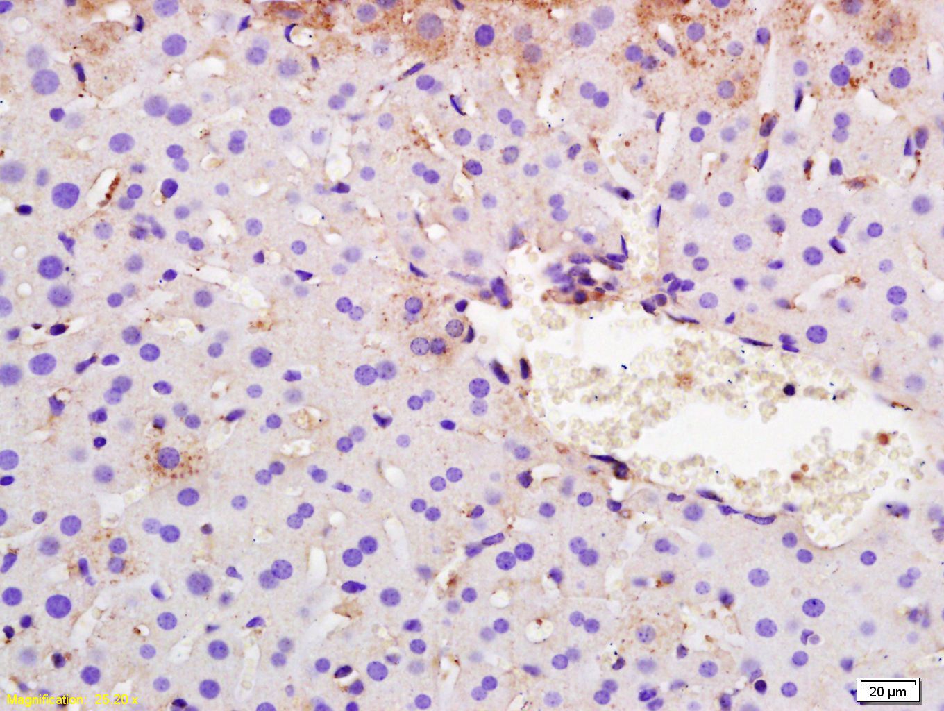
Fig1: Tissue/cell: mouse liver tissue; 4% Paraformaldehyde-fixed and paraffin-embedded; Antigen retrieval: citrate buffer ( 0.01M, pH 6.0 ), Boiling bathing for 15min; Block endogenous peroxidase by 3% Hydrogen peroxide for 30min; Blocking buffer (normal goat serum,C-0005) at 37℃ for 20 min; Incubation: Anti-SYVN1/HRD1 Polyclonal Antibody, Unconjugated(ER1917-45) 1:200, overnight at 4℃, followed by conjugation to the secondary antibody(SP-0023) and DAB(C-0010) stainingApplication
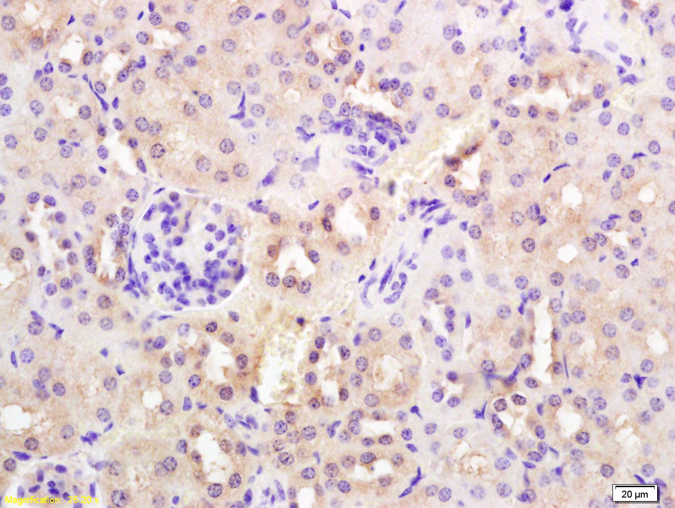
Fig2: Tissue/cell: mouse kidney tissue; 4% Paraformaldehyde-fixed and paraffin-embedded; Antigen retrieval: citrate buffer ( 0.01M, pH 6.0 ), Boiling bathing for 15min; Block endogenous peroxidase by 3% Hydrogen peroxide for 30min; Blocking buffer (normal goat serum,C-0005) at 37℃ for 20 min; Incubation: Anti-SYVN1/HRD1 Polyclonal Antibody, Unconjugated(ER1917-45) 1:200, overnight at 4℃, followed by conjugation to the secondary antibody(SP-0023) and DAB(C-0010) stainingApplication
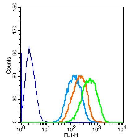
Fig3: The figure annotation: The blue histogram is unstained cells. The Wathet Blue histogram is cells stained with secondary antibody (bs-0295G-FITC) alone. The Orange histogram is cells stained with rabbit IgG isotype control antibody(bs-0295P)plus secondary antibody. The green histogram is cells stained with Rabbit Anti-SYVN1 antibody (ER1917-45) plus secondary antibody. Positive control: Hepg2 cells Concentration: 1:50Application
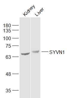
Fig4: Sample: Kidney (Mouse) Lysate at 40 ug Liver (Mouse) Lysate at 40 ug Primary: Anti-SYVN1 (ER1917-45) at 1/1000 dilution Secondary: IRDye800CW Goat Anti-Rabbit IgG at 1/20000 dilution Predicted band size: 65 kD Observed band size: 65/70 kD| Product Name | SYVN1 |
|---|---|
| Antibody Type | Primary Antibodies |
| Antigen Alias | 1200010C09Rik HRD1 KIAA1810 MGC40372 Synovial apoptosis inhibitor 1 synoviolin Synoviolin 1 isoform b SYVN1_HUMAN DER3 E3 ubiquitin-protein ligase synoviolin HMG coA reductase degradation 1 homolog OTTHUMP00000230429 OTTHUMP00000230430 OTTHUMP00000230431 OTTHUMP00000230432 Synovial apoptosis inhibitor 1 Synoviolin 1 isoform b SYNOVIOLIN SYVN1. |
| Product description | Acts as an E3 ubiquitin-protein ligase which accepts ubiquitin specifically from endoplasmic reticulum-associated UBC7 E2 ligase and transfers it to substrates, promoting their degradation. Component of the endoplasmic reticulum quality control (ERQC) system also called ER-associated degradation (ERAD) involved in ubiquitin-dependent degradation of misfolded endoplasmic reticulum proteins. Also promotes the degradation of normal but naturally short-lived proteins such as SGK. Protects cells from ER stress-induced apoptosis. Protects neurons from apoptosis induced by polyglutamine-expanded huntingtin (HTT) or unfolded GPR37 by promoting their degradation. Sequesters p53/TP53 in the cytoplasm and promotes its degradation, thereby negatively regulating its biological function in transcription, cell cycle regulation and apoptosis. |
| Immunogen | KLH conjugated synthetic peptide derived from human SYVN1 531-617/617 |
| Clonality | Polyclonal |
|---|---|
| Isotype | IgG |
| Host Species | Rabbit |
| Tested Applications | WBIHC-PFC |
| WB:1:500-2000: IHC-P:1:400-800: FC:3μg/Test: |
|
| Species Reactivity | HumanMouseRatDogCow |
| Concentration | 1mg/ml |
| Alternative Names | 1200010C09Rik HRD1 KIAA1810 MGC40372 Synovial apoptosis inhibitor 1 synoviolin Synoviolin 1 isoform b SYVN1_HUMAN DER3 E3 ubiquitin-protein ligase synoviolin HMG coA reductase degradation 1 homolog OTTHUMP00000230429 OTTHUMP00000230430 OTTHUMP00000230431 OTTHUMP00000230432 Synovial apoptosis inhibitor 1 Synoviolin 1 isoform b SYNOVIOLIN SYVN1. |
|---|---|
| Molecular Weight(MW) | 65 KDa |
| Cellular Localization | Endoplasmic reticulum membrane; Multi-pass membrane protein. |
| SwissProt ID | Q86TM6 |
|---|
Application
Fig1: Tissue/cell: mouse liver tissue; 4% Paraformaldehyde-fixed and paraffin-embedded; Antigen retrieval: citrate buffer ( 0.01M, pH 6.0 ), Boiling bathing for 15min; Block endogenous peroxidase by 3% Hydrogen peroxide for 30min; Blocking buffer (normal goat serum,C-0005) at 37℃ for 20 min; Incubation: Anti-SYVN1/HRD1 Polyclonal Antibody, Unconjugated(ER1917-45) 1:200, overnight at 4℃, followed by conjugation to the secondary antibody(SP-0023) and DAB(C-0010) staining
Application
Fig2: Tissue/cell: mouse kidney tissue; 4% Paraformaldehyde-fixed and paraffin-embedded; Antigen retrieval: citrate buffer ( 0.01M, pH 6.0 ), Boiling bathing for 15min; Block endogenous peroxidase by 3% Hydrogen peroxide for 30min; Blocking buffer (normal goat serum,C-0005) at 37℃ for 20 min; Incubation: Anti-SYVN1/HRD1 Polyclonal Antibody, Unconjugated(ER1917-45) 1:200, overnight at 4℃, followed by conjugation to the secondary antibody(SP-0023) and DAB(C-0010) staining
Application
Fig3: The figure annotation: The blue histogram is unstained cells. The Wathet Blue histogram is cells stained with secondary antibody (bs-0295G-FITC) alone. The Orange histogram is cells stained with rabbit IgG isotype control antibody(bs-0295P)plus secondary antibody. The green histogram is cells stained with Rabbit Anti-SYVN1 antibody (ER1917-45) plus secondary antibody. Positive control: Hepg2 cells Concentration: 1:50
Application
Fig4: Sample: Kidney (Mouse) Lysate at 40 ug Liver (Mouse) Lysate at 40 ug Primary: Anti-SYVN1 (ER1917-45) at 1/1000 dilution Secondary: IRDye800CW Goat Anti-Rabbit IgG at 1/20000 dilution Predicted band size: 65 kD Observed band size: 65/70 kD| Positive Control | Ubiquitously expressed, with highest levels in liver and kidney (at protein level). Up-regulated in synovial tissues from patients with rheumatoid arthritis (at protein level). |
|---|---|
| Application Notes | WB:1:500-2000: IHC-P:1:400-800: FC:3μg/Test: |
| Form | Liquid |
|---|---|
| Storage Instructions | Store at -20℃ for one year. Avoid repeated freeze/thaw cycles. The lyophilized antibody is stable at room temperature for at least one month and for greater than a year when kept at -20℃. When reconstituted in sterile pH 7.4 0.01M PBS or diluent of antibody the antibody is stable for at least two weeks at 2-4℃. |
| Storage Buffer | 0.01M TBS(pH7.4) with 1% BSA, 0.03% Proclin300 and 50% Glycerol. |


