Application
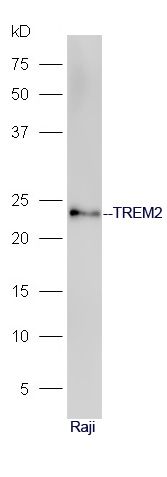
Fig1: Sample: Raji Cell lysate at 30ug; Primary: Anti-TREM2(ER1918-04) at 1:300 dilution; Secondary: HRP conjugated Goat-Anti-rabbit IgG(bs-0295G-HRP) at 1: 5000 dilution; Predicted band size: 23 kD Observed band size: 23 kDApplication
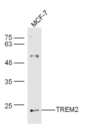
Fig2: Sample: MCF-7 Cell (Human) Lysate at 30 ug Primary: Anti-TREM2 (ER1918-04) at 1/300 dilution Secondary: IRDye800CW Goat Anti-Rabbit IgG at 1/20000 dilution Predicted band size: 23 kD Observed band size: 23 kDApplication
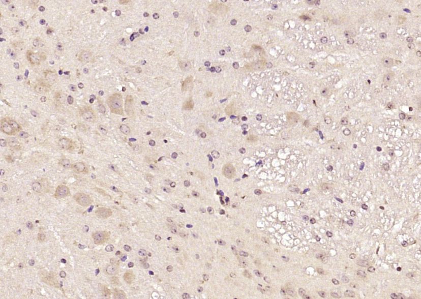
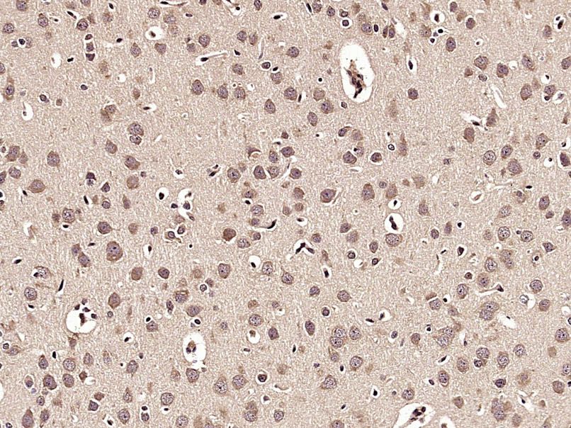
Fig3: Paraformaldehyde-fixed, paraffin embedded (mouse brain); Antigen retrieval by boiling in sodium citrate buffer (pH6.0) for 15min; Block endogenous peroxidase by 3% hydrogen peroxide for 20 minutes; Blocking buffer (normal goat serum) at 37℃ for 30min; Antibody incubation with (TREM2) Polyclonal Antibody, Unconjugated (ER1918-04) at 1:200 overnight at 4℃, followed by operating according to SP Kit(Rabbit) (sp-0023) instructionsand DAB staining.Application
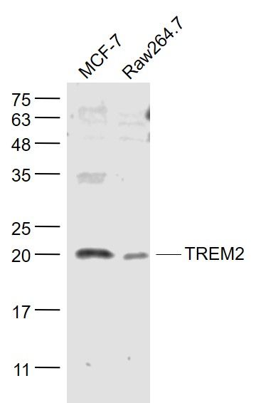
Fig4: Sample: MCF-7(Human) Cell Lysate at 30 ug Raw264.7(Mouse) Cell Lysate at 30 ug Primary: Anti- TREM2 (ER1918-04) at 1/1000 dilution Secondary: IRDye800CW Goat Anti-Rabbit IgG at 1/20000 dilution Predicted band size: 23 kD Observed band size: 20 kDApplication
Fig5: Paraformaldehyde-fixed, paraffin embedded (Rat brain); Antigen retrieval by microwave in sodium citrate buffer (pH6.0) ; Block endogenous peroxidase by 3% hydrogen peroxide for 30 minutes; Blocking buffer (3% BSA) at RT for 30min; Antibody incubation with (TREM2) Polyclonal Antibody, Unconjugated (ER1918-04) at 1:400 overnight at 4℃, followed by conjugation to the secondary antibody (labeled with HRP)and DAB staining.| Product Name | TREM2 |
|---|---|
| Antibody Type | Primary Antibodies |
| Antigen Alias | TREM-2 TREM2a TREM2b TREM2c Trggering receptor expressed on myeloid cells 2 Trggering receptor expressed on myeloid cells 2a Triggering receptor expressed on monocytes 2 TREM2_HUMAN. |
| Product description | Forms a receptor signaling complex with TYROBP which mediates signaling and cell activation following ligand binding. Acts as a receptor for amyloid-beta protein 42, a cleavage product of the amyloid-beta precursor protein APP, and mediates its uptake and degradation by microglia. Binding to amyloid-beta 42 mediates microglial activation, proliferation, migration, apoptosis and expression of pro-inflammatory cytokines, such as IL6R and CCL3, and the anti-inflammatory cytokine ARG1. Acts as a receptor for lipoprotein particles such as LDL, VLDL, and HDL and for apolipoproteins such as APOA1, APOA2, APOB, APOE, APOE2, APOE3, APOE4, and CLU and enhances their uptake in microglia. Binds phospholipids (preferably anionic lipids) such as phosphatidylserine, phosphatidylethanolamine, phosphatidylglycerol and sphingomyelin. Regulates microglial proliferation by acting as an upstream regulator of the Wnt/beta-catenin signaling cascade. Required for microglial phagocytosis of apoptotic neurons. Also required for microglial activation and phagocytosis of myelin debris after neuronal injury and of neuronal synapses during synapse elimination in the developing brain. |
| Immunogen | KLH conjugated synthetic peptide derived from human TREM2 1-100/230 |
| Clonality | Polyclonal |
|---|---|
| Isotype | IgG |
| Host Species | Rabbit |
| Tested Applications | WBIHC-P |
| WB:1:500-2000: IHC-P:1:400-800: |
|
| Species Reactivity | HumanMouseRatDogPigCowHorse |
| Concentration | 1mg/ml |
| Alternative Names | TREM-2
TREM2a
TREM2b TREM2c Trggering receptor expressed on myeloid cells 2 Trggering receptor expressed on myeloid cells 2a Triggering receptor expressed on monocytes 2 TREM2_HUMAN. |
|---|---|
| Molecular Weight(MW) | 25 KDa |
| Cellular Localization | Isoform 1: Cell membrane; Single-pass type I membrane protein (Potential). Isoform 2: Secreted (Potential). Isoform 3: Secreted (Potential). |
| SwissProt ID | Q9NZC2 |
|---|
Application
Fig1: Sample: Raji Cell lysate at 30ug; Primary: Anti-TREM2(ER1918-04) at 1:300 dilution; Secondary: HRP conjugated Goat-Anti-rabbit IgG(bs-0295G-HRP) at 1: 5000 dilution; Predicted band size: 23 kD Observed band size: 23 kD
Application
Fig2: Sample: MCF-7 Cell (Human) Lysate at 30 ug Primary: Anti-TREM2 (ER1918-04) at 1/300 dilution Secondary: IRDye800CW Goat Anti-Rabbit IgG at 1/20000 dilution Predicted band size: 23 kD Observed band size: 23 kD
Application
Fig3: Paraformaldehyde-fixed, paraffin embedded (mouse brain); Antigen retrieval by boiling in sodium citrate buffer (pH6.0) for 15min; Block endogenous peroxidase by 3% hydrogen peroxide for 20 minutes; Blocking buffer (normal goat serum) at 37℃ for 30min; Antibody incubation with (TREM2) Polyclonal Antibody, Unconjugated (ER1918-04) at 1:200 overnight at 4℃, followed by operating according to SP Kit(Rabbit) (sp-0023) instructionsand DAB staining.
Application
Fig4: Sample: MCF-7(Human) Cell Lysate at 30 ug Raw264.7(Mouse) Cell Lysate at 30 ug Primary: Anti- TREM2 (ER1918-04) at 1/1000 dilution Secondary: IRDye800CW Goat Anti-Rabbit IgG at 1/20000 dilution Predicted band size: 23 kD Observed band size: 20 kD
Application
Fig5: Paraformaldehyde-fixed, paraffin embedded (Rat brain); Antigen retrieval by microwave in sodium citrate buffer (pH6.0) ; Block endogenous peroxidase by 3% hydrogen peroxide for 30 minutes; Blocking buffer (3% BSA) at RT for 30min; Antibody incubation with (TREM2) Polyclonal Antibody, Unconjugated (ER1918-04) at 1:400 overnight at 4℃, followed by conjugation to the secondary antibody (labeled with HRP)and DAB staining.| Positive Control | Expressed on macrophages and dendritic cells but not on granulocytes or monocytes. In the CNS strongest expression seen in the basal ganglia, corpus callosum, medulla oblongata and spinal cord. |
|---|---|
| Application Notes | WB:1:500-2000: IHC-P:1:400-800: |
| Form | Liquid |
|---|---|
| Storage Instructions | Store at -20℃ for one year. Avoid repeated freeze/thaw cycles. |
| Storage Buffer | 0.01M TBS(pH7.4) with 1% BSA, 0.03% Proclin300 and 50% Glycerol. |


