Application
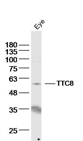
Fig1: Sample: eye (mouse) Lysate at 40 ug Primary: Anti- TTC8 (ER1918-27)at 1/300 dilution Secondary: IRDye800CW Goat Anti-Rabbit IgG at 1/20000 dilution Predicted band size: 61kD Observed band size: 58 kDApplication
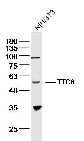
Fig2: Sample: NIH/3T3 (human)cell Lysate at 40 ug Primary: Anti- TTC8 (ER1918-27)at 1/300 dilution Secondary: IRDye800CW Goat Anti-Rabbit IgG at 1/20000 dilution Predicted band size: 61kD Observed band size: 58 kDApplication
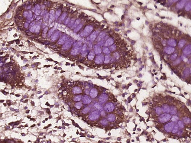
Fig3: Paraformaldehyde-fixed, paraffin embedded (Human colon cancer); Antigen retrieval by boiling in sodium citrate buffer (pH6.0) for 15min; Block endogenous peroxidase by 3% hydrogen peroxide for 20 minutes; Blocking buffer (normal goat serum) at 37℃ for 30min; Antibody incubation with (TTC8) Polyclonal Antibody, Unconjugated (ER1918-27) at 1:400 overnight at 4℃, followed by operating according to SP Kit(Rabbit) (sp-0023) instructionsand DAB staining.Application
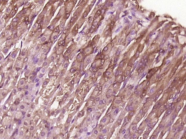
Fig4: Paraformaldehyde-fixed, paraffin embedded (Mouse stomach); Antigen retrieval by boiling in sodium citrate buffer (pH6.0) for 15min; Block endogenous peroxidase by 3% hydrogen peroxide for 20 minutes; Blocking buffer (normal goat serum) at 37℃ for 30min; Antibody incubation with (TTC8) Polyclonal Antibody, Unconjugated (ER1918-27) at 1:400 overnight at 4℃, followed by operating according to SP Kit(Rabbit) (sp-0023) instructionsand DAB staining.Application
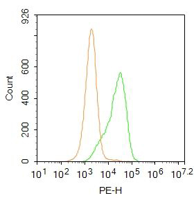
Fig5: Blank control:A549. Primary Antibody (green line): Rabbit Anti-TTC8 antibody (ER1918-27) Dilution: 1μg /10^6 cells; Isotype Control Antibody (orange line): Rabbit IgG . Secondary Antibody : Goat anti-rabbit IgG-PE Dilution:0.2μg /test. Protocol The cells were fixed with 4% PFA (10min at room temperature)and then permeabilized with 20% PBST for 20 min at room temperature. The cells were then incubated in 5% BSA to block non-specific protein-protein interactions for 30 min at at room temperature .Cells stained with Primary Antibody for 30 min at room temperature. The secondary antibody used for 40 min at room temperature. Acquisition of 20,000 events was performed.| Product Name | TTC8 |
|---|---|
| Antibody Type | Primary Antibodies |
| Antigen Alias | Bardet Biedl syndrome 8 protein Bardet Biedl syndrome type 8 Bardet-Biedl syndrome 8 protein BBS8 Tetratricopeptide repeat domain 8 Tetratricopeptide repeat protein 8 TPR repeat protein 8 TTC 8 Ttc8 TTC8_HUMAN. |
| Product description | The BBSome complex is required for ciliogenesis but is dispensable for centriolar satellite function. This ciliogenic function is mediated in part by the Rab8 GDP/GTP exchange factor, which localizes to the basal body and contacts the BBSome. Rab8(GTP) enters the primary cilium and promotes extension of the ciliary membrane. Firstly the BBSome associates with the ciliary membrane and binds to RAB3IP/Rabin8, the guanosyl exchange factor (GEF) for Rab8 and then the Rab8-GTP localizes to the cilium and promotes docking and fusion of carrier vesicles to the base of the ciliary membrane. |
| Immunogen | KLH conjugated synthetic peptide derived from human BBS8 251-330/541 |
| Clonality | Polyclonal |
|---|---|
| Isotype | IgG |
| Host Species | Rabbit |
| Tested Applications | WBIHC-PFC |
| WB:1:500-2000: IHC-P:1:400-800: FC:0.2ug/test: |
|
| Species Reactivity | HumanMouseRatChickenDogPigCowHorseRabbitSheep |
| Concentration | 1mg/ml |
| Alternative Names | Bardet Biedl syndrome 8 protein Bardet Biedl syndrome type 8 Bardet-Biedl syndrome 8 protein BBS8 Tetratricopeptide repeat domain 8 Tetratricopeptide repeat protein 8 TPR repeat protein 8 TTC 8 Ttc8 TTC8_HUMAN. |
|---|---|
| Molecular Weight(MW) | 61 KDa |
| Cellular Localization | Cytoplasm, cytoskeleton, centrosome. Cell projection, cilium membrane. Cytoplasm. Note=Localizes to nonmembranous centriolar satellites in the cytoplasm. |
| SwissProt ID | Q8TAM2 |
|---|
Application
Fig1: Sample: eye (mouse) Lysate at 40 ug Primary: Anti- TTC8 (ER1918-27)at 1/300 dilution Secondary: IRDye800CW Goat Anti-Rabbit IgG at 1/20000 dilution Predicted band size: 61kD Observed band size: 58 kD
Application
Fig2: Sample: NIH/3T3 (human)cell Lysate at 40 ug Primary: Anti- TTC8 (ER1918-27)at 1/300 dilution Secondary: IRDye800CW Goat Anti-Rabbit IgG at 1/20000 dilution Predicted band size: 61kD Observed band size: 58 kD
Application
Fig3: Paraformaldehyde-fixed, paraffin embedded (Human colon cancer); Antigen retrieval by boiling in sodium citrate buffer (pH6.0) for 15min; Block endogenous peroxidase by 3% hydrogen peroxide for 20 minutes; Blocking buffer (normal goat serum) at 37℃ for 30min; Antibody incubation with (TTC8) Polyclonal Antibody, Unconjugated (ER1918-27) at 1:400 overnight at 4℃, followed by operating according to SP Kit(Rabbit) (sp-0023) instructionsand DAB staining.
Application
Fig4: Paraformaldehyde-fixed, paraffin embedded (Mouse stomach); Antigen retrieval by boiling in sodium citrate buffer (pH6.0) for 15min; Block endogenous peroxidase by 3% hydrogen peroxide for 20 minutes; Blocking buffer (normal goat serum) at 37℃ for 30min; Antibody incubation with (TTC8) Polyclonal Antibody, Unconjugated (ER1918-27) at 1:400 overnight at 4℃, followed by operating according to SP Kit(Rabbit) (sp-0023) instructionsand DAB staining.
Application
Fig5: Blank control:A549. Primary Antibody (green line): Rabbit Anti-TTC8 antibody (ER1918-27) Dilution: 1μg /10^6 cells; Isotype Control Antibody (orange line): Rabbit IgG . Secondary Antibody : Goat anti-rabbit IgG-PE Dilution:0.2μg /test. Protocol The cells were fixed with 4% PFA (10min at room temperature)and then permeabilized with 20% PBST for 20 min at room temperature. The cells were then incubated in 5% BSA to block non-specific protein-protein interactions for 30 min at at room temperature .Cells stained with Primary Antibody for 30 min at room temperature. The secondary antibody used for 40 min at room temperature. Acquisition of 20,000 events was performed.| Positive Control | Widely expressed. |
|---|---|
| Application Notes | WB:1:500-2000: IHC-P:1:400-800: FC:0.2ug/test: |
| Form | Liquid |
|---|---|
| Storage Instructions | Store at -20℃ for one year. Avoid repeated freeze/thaw cycles. The lyophilized antibody is stable at room temperature for at least one month and for greater than a year when kept at -20℃. When reconstituted in sterile pH 7.4 0.01M PBS or diluent of antibody the antibody is stable for at least two weeks at 2-4℃. |
| Storage Buffer | 0.01M TBS(pH7.4) with 1% BSA, 0.03% Proclin300 and 50% Glycerol. |


