Application
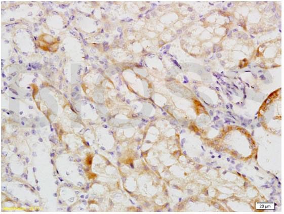
Fig1: Tissue/cell: mouse kidney tissue; 4% Paraformaldehyde-fixed and paraffin-embedded; Antigen retrieval: citrate buffer ( 0.01M, pH 6.0 ), Boiling bathing for 15min; Block endogenous peroxidase by 3% Hydrogen peroxide for 30min; Blocking buffer (normal goat serum,C-0005) at 37℃ for 20 min; Incubation: Anti-MCKD2/UMOD Polyclonal Antibody, Unconjugated(ER1918-44) 1:200, overnight at 4℃, followed by conjugation to the secondary antibody(SP-0023) and DAB(C-0010) stainingApplication
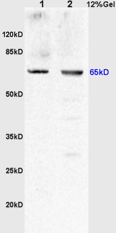
Fig2: Sample: Lane 1: Kidney(Rat) Lysate at 30 ug Lane 2: Kidney carcinoma(Human) Lysate at 30 ug Primary: Anti-MCKD2/UMOD (ER1918-44) at 1:200 dilution; Secondary: HRP conjugated Goat Anti-Rabbit IgG(bs-0295G-HRP) at 1: 3000 dilution; Predicted band size : 65kD Observed band size : 65kDApplication
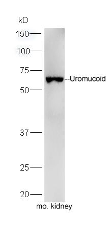
Fig3: Sample: Kidney(Mouse) lysate at 30 ug; Primary: Anti-Uromucoid (ER1918-44) at 1:300 dilution; Secondary: HRP conjugated Goat-Anti-rabbit IgG(bs-0295G-HRP) at 1: 5000 dilution; Predicted band size:61/65 kD Observed band size:65 kDApplication
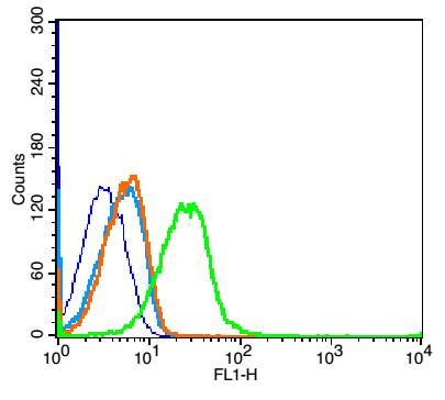
Fig4: Blank control: Mouse kidney (blue). Primary Antibody:Rabbit Anti-Coxsackie Adenovirus Receptor antibody (ER1918-44,Green); Dilution: 1μg in 100 μL 1X PBS containing 0.5% BSA; Isotype Control Antibody: Rabbit IgG(orange) ,used under the same conditions; Secondary Antibody: Goat anti-rabbit IgG-FITC(white blue), Dilution: 1:200 in 1 X PBS containing 0.5% BSA. Protocol The cells were fixed with 2% paraformaldehyde for 10 min at 37℃. Primary antibody (ER1918-44, 1μg /1x10^6 cells) were incubated for 30 min at room temperature, followed by 1 X PBS containing 0.5% BSA + 10% goat serum (1 hour) to block non-specific protein-protein interactions. Then the Goat Anti-rabbit IgG/FITC antibody was added into the blocking buffer mentioned above to react with the primary antibody at 1/200 dilution for 40 min at room temperature. Acquisition of 20,000 events was performed.| Product Name | Uromucoid |
|---|---|
| Antibody Type | Primary Antibodies |
| Antigen Alias | UMOD ADMCKD2 FJHN HNFJ HNFJ1 MCKD2 medullary cystic kidney disease 2 (autosomal dominant) Tamm Horsfall glycoprotein Tamm Horsfall urinary glycoprotein Tamm-Horsfall urinary glycoprotein THGP THP Umod UROM_MOUSE uromodulin (uromucoid, Tamm-Horsfall glycoprotein) Uromodulin Uromodulin, secreted form. |
| Product description | Uromodulin: Functions in biogenesis and organization of the apical membrane of epithelial cells of the thick ascending limb of Henle's loop (TALH), where it promotes formation of complex filamentous gel-like structure providing the water barrier permeability. May serve as a receptor for binding and endocytosis for cytokines (IL-1, IL-2) and TNF. Facilitates neutrophil migration across renal epithelial (By similarity).;Uromodulin, secreted form: Secreted into urine after proteolytically cleaveage. Into the urine, may contribute to colloid osmotic pressure, retards passage of positively charged electrolytes, prevents urinary tract infection and modulates formation of supersaturated salts and their crystals. |
| Immunogen | KLH conjugated synthetic peptide derived from mouse MCKD2 351-450/642 |
| Clonality | Polyclonal |
|---|---|
| Isotype | IgG |
| Host Species | Rabbit |
| Tested Applications | WBIHC-PICC/IFFC |
| WB:1:500-2000: IHC-P:1:400-800: FC:1: |
|
| Species Reactivity | HumanMouseRatCow |
| Concentration | 1mg/ml |
| Alternative Names | UMOD
ADMCKD2
FJHN
HNFJ
HNFJ1
MCKD2
medullary cystic kidney disease 2 (autosomal dominant)
Tamm Horsfall glycoprotein
Tamm Horsfall urinary glycoprotein
Tamm-Horsfall urinary glycoprotein
THGP
THP
Umod
UROM_MOUSE
uromodulin (uromucoid Tamm-Horsfall glycoprotein) Uromodulin Uromodulin secreted form. |
|---|---|
| Molecular Weight(MW) | 61/65 KDa |
| Cellular Localization | Apical cell membrane; Lipid-anchor, GPI-anchor (By similarity). Basolateral cell membrane; Lipid-anchor, GPI-anchor (By similarity). Cell projection, cilium membrane (By similarity). Note=Only a small fraction is sorts to the basolateral pole of tubular epithelial cells compared to apical localization (By similarity). Uromodulin, secreted form: Secreted (By similarity). |
| SwissProt ID | Q91X17 |
|---|
Application
Fig1: Tissue/cell: mouse kidney tissue; 4% Paraformaldehyde-fixed and paraffin-embedded; Antigen retrieval: citrate buffer ( 0.01M, pH 6.0 ), Boiling bathing for 15min; Block endogenous peroxidase by 3% Hydrogen peroxide for 30min; Blocking buffer (normal goat serum,C-0005) at 37℃ for 20 min; Incubation: Anti-MCKD2/UMOD Polyclonal Antibody, Unconjugated(ER1918-44) 1:200, overnight at 4℃, followed by conjugation to the secondary antibody(SP-0023) and DAB(C-0010) staining
Application
Fig2: Sample: Lane 1: Kidney(Rat) Lysate at 30 ug Lane 2: Kidney carcinoma(Human) Lysate at 30 ug Primary: Anti-MCKD2/UMOD (ER1918-44) at 1:200 dilution; Secondary: HRP conjugated Goat Anti-Rabbit IgG(bs-0295G-HRP) at 1: 3000 dilution; Predicted band size : 65kD Observed band size : 65kD
Application
Fig3: Sample: Kidney(Mouse) lysate at 30 ug; Primary: Anti-Uromucoid (ER1918-44) at 1:300 dilution; Secondary: HRP conjugated Goat-Anti-rabbit IgG(bs-0295G-HRP) at 1: 5000 dilution; Predicted band size:61/65 kD Observed band size:65 kD
Application
Fig4: Blank control: Mouse kidney (blue). Primary Antibody:Rabbit Anti-Coxsackie Adenovirus Receptor antibody (ER1918-44,Green); Dilution: 1μg in 100 μL 1X PBS containing 0.5% BSA; Isotype Control Antibody: Rabbit IgG(orange) ,used under the same conditions; Secondary Antibody: Goat anti-rabbit IgG-FITC(white blue), Dilution: 1:200 in 1 X PBS containing 0.5% BSA. Protocol The cells were fixed with 2% paraformaldehyde for 10 min at 37℃. Primary antibody (ER1918-44, 1μg /1x10^6 cells) were incubated for 30 min at room temperature, followed by 1 X PBS containing 0.5% BSA + 10% goat serum (1 hour) to block non-specific protein-protein interactions. Then the Goat Anti-rabbit IgG/FITC antibody was added into the blocking buffer mentioned above to react with the primary antibody at 1/200 dilution for 40 min at room temperature. Acquisition of 20,000 events was performed.| Positive Control | Mouse kidney tissue, Kidney(Rat) Lysate, Kidney carcinoma(Human) Lysate, Kidney(Mouse) lysate. |
|---|---|
| Application Notes | WB:1:500-2000: IHC-P:1:400-800: FC:1: |
| Form | Liquid |
|---|---|
| Storage Instructions | Store at -20℃ for one year. Avoid repeated freeze/thaw cycles. The lyophilized antibody is stable at room temperature for at least one month and for greater than a year when kept at -20℃. When reconstituted in sterile pH 7.4 0.01M PBS or diluent of antibody the antibody is stable for at least two weeks at 2-4℃. |
| Storage Buffer | 0.01M TBS(pH7.4) with 1% BSA, 0.03% Proclin300 and 50% Glycerol. |


