Application
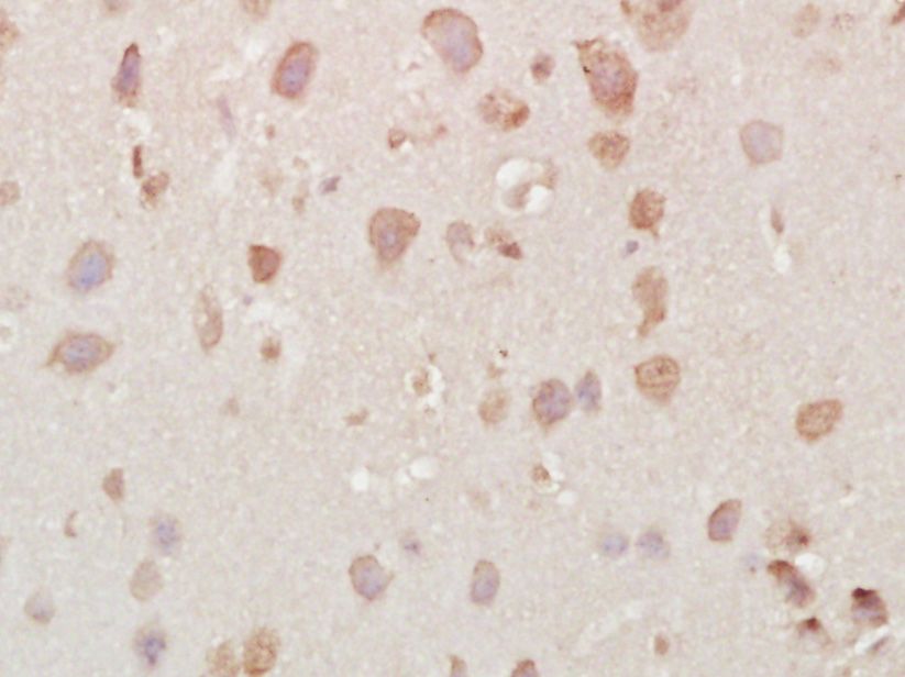
Fig1: Paraformaldehyde-fixed, paraffin embedded (Mouse brain); Antigen retrieval by boiling in sodium citrate buffer (pH6.0) for 15min; Block endogenous peroxidase by 3% hydrogen peroxide for 20 minutes; Blocking buffer (normal goat serum) at 37℃ for 30min; Antibody incubation with (Vesicle docking protein p115) Polyclonal Antibody, Unconjugated (ER1918-51) at 1:400 overnight at 4℃, followed by operating according to SP Kit(Rabbit) (sp-0023) instructionsand DAB staining.Application
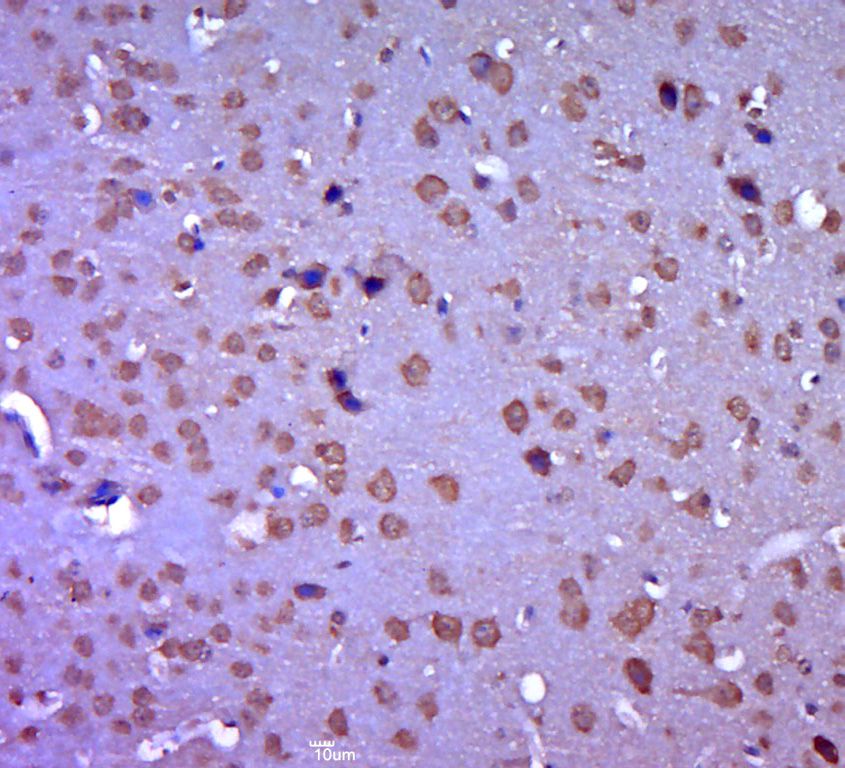
Fig2: Paraformaldehyde-fixed, paraffin embedded (mouse brain tissue); Antigen retrieval by boiling in sodium citrate buffer (pH6.0) for 15min; Block endogenous peroxidase by 3% hydrogen peroxide for 20 minutes; Blocking buffer (normal goat serum) at 37℃ for 30min; Antibody incubation with (Vesicle docking protein p115) Polyclonal Antibody, Unconjugated (ER1918-51) at 1:400 overnight at 4℃, followed by a conjugated secondary (sp-0023) for 20 minutes and DAB staining.Application
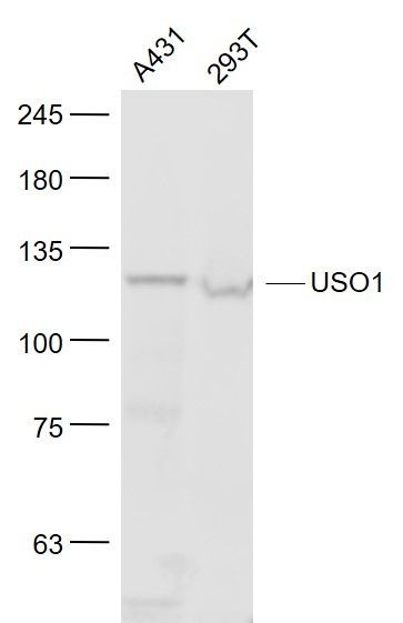
Fig3: Sample: A431(Human) Cell Lysate at 30 ug 293T(Human) Cell Lysate at 30 ug Primary: Anti- USO1 (ER1918-51) at 1/1000 dilution Secondary: IRDye800CW Goat Anti-Rabbit IgG at 1/20000 dilution Predicted band size: 108 kD Observed band size: 120 kDApplication
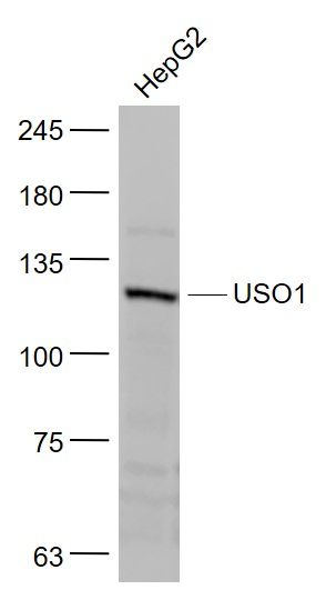
Fig4: Sample: HepG2(Human) Cell Lysate at 30 ug Primary: Anti- USO1 (ER1918-51) at 1/1000 dilution Secondary: IRDye800CW Goat Anti-Rabbit IgG at 1/20000 dilution Predicted band size: 108 kD Observed band size: 120 kD| Product Name | Vesicle docking protein p115 |
|---|---|
| Antibody Type | Primary Antibodies |
| Antigen Alias | P115-RhoGEF General vesicular transport factor General vesicular transport factor p115 P115 TAP Transcytosis associated protein VDP Vesicle docking protein USO1_HUMAN. |
| Product description | General vesicular transport factor required for intercisternal transport in the Golgi stack; it is required for transcytotic fusion and/or subsequent binding of the vesicles to the target membrane. May well act as a vesicular anchor by interacting with the target membrane and holding the vesicular and target membranes in proximity. |
| Immunogen | KLH conjugated synthetic peptide derived from human Vesicle docking protein p115 501-600/962 |
| Clonality | Polyclonal |
|---|---|
| Isotype | IgG |
| Host Species | Rabbit |
| Tested Applications | WBIHC-P |
| WB:1:500-2000: IHC-P:1:400-800: |
|
| Species Reactivity | HumanMouseRatChickenPigCowHorseSheep |
| Concentration | 1mg/ml |
| Alternative Names | P115-RhoGEF General vesicular transport factor General vesicular transport factor p115 P115 TAP Transcytosis associated protein VDP Vesicle docking protein USO1_HUMAN. |
|---|---|
| Molecular Weight(MW) | 108 KDa |
| Cellular Localization | Cytoplasm; cytosol. Golgi apparatus membrane. Recycles between the cytosol and the Golgi apparatus during interphase. During interphase, the phosphorylated form is found exclusively in cytosol; the unphosphorylated form is associated with Golgi apparatus membranes. |
| SwissProt ID | O60763 |
|---|
Application
Fig1: Paraformaldehyde-fixed, paraffin embedded (Mouse brain); Antigen retrieval by boiling in sodium citrate buffer (pH6.0) for 15min; Block endogenous peroxidase by 3% hydrogen peroxide for 20 minutes; Blocking buffer (normal goat serum) at 37℃ for 30min; Antibody incubation with (Vesicle docking protein p115) Polyclonal Antibody, Unconjugated (ER1918-51) at 1:400 overnight at 4℃, followed by operating according to SP Kit(Rabbit) (sp-0023) instructionsand DAB staining.
Application
Fig2: Paraformaldehyde-fixed, paraffin embedded (mouse brain tissue); Antigen retrieval by boiling in sodium citrate buffer (pH6.0) for 15min; Block endogenous peroxidase by 3% hydrogen peroxide for 20 minutes; Blocking buffer (normal goat serum) at 37℃ for 30min; Antibody incubation with (Vesicle docking protein p115) Polyclonal Antibody, Unconjugated (ER1918-51) at 1:400 overnight at 4℃, followed by a conjugated secondary (sp-0023) for 20 minutes and DAB staining.
Application
Fig3: Sample: A431(Human) Cell Lysate at 30 ug 293T(Human) Cell Lysate at 30 ug Primary: Anti- USO1 (ER1918-51) at 1/1000 dilution Secondary: IRDye800CW Goat Anti-Rabbit IgG at 1/20000 dilution Predicted band size: 108 kD Observed band size: 120 kD
Application
Fig4: Sample: HepG2(Human) Cell Lysate at 30 ug Primary: Anti- USO1 (ER1918-51) at 1/1000 dilution Secondary: IRDye800CW Goat Anti-Rabbit IgG at 1/20000 dilution Predicted band size: 108 kD Observed band size: 120 kD| Application Notes | WB:1:500-2000: IHC-P:1:400-800: |
|---|
| Form | Liquid |
|---|---|
| Storage Instructions | Store at -20℃ for one year. Avoid repeated freeze/thaw cycles. The lyophilized antibody is stable at room temperature for at least one month and for greater than a year when kept at -20℃. When reconstituted in sterile pH 7.4 0.01M PBS or diluent of antibody the antibody is stable for at least two weeks at 2-4℃. |
| Storage Buffer | 0.01M TBS(pH7.4) with 1% BSA, 0.03% Proclin300 and 50% Glycerol. |


