Application
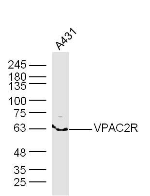
Fig1: Sample: A431 Cell Lysate at 40 ug Primary: Anti- VPAC2R (ER1918-56) at 1/300 dilution Secondary: IRDye800CW Goat Anti-Rabbit IgG at 1/20000 dilution Predicted band size: 48 kD Observed band size: 63 kD| Product Name | VPAC2R |
|---|---|
| Antibody Type | Primary Antibodies |
| Antigen Alias | VIP receptor II VIP receptor2 VIP receptor-2 PACAP type-3 VIP type-2 receptor Vipr2 Vip2 VPAC2 VPAC2R Vasoactive intestinal polypeptide receptor 2 VIP-R-2 Pituitary adenylate cyclase-activating polypeptide type III receptor PACAP type III receptor PACAP-R-3 PACAP-R3 VIPR2_HUMAN. |
| Product description | ng polypeptide (PACAP) to some degree. Both receptors are members of the 7 transmembrane G protein-coupled receptor family. VPAC1 is distributed widely in the CNS, liver, lung, intestine and T-lymphocytes. VPAC2 is found in the CNS, pancreas, skeletal muscle, heart, kidney, adipose tissue, testis, and stomach. Vasoactive intestinal peptide (VIP) and pituitary adenylate cyclase-activating peptide (PACAP) receptors are activated by the endogenous peptides VIP, PACAP-38, PACAP-27, peptide histidine isoleucineamide (PHI), peptide histidine methionineamide (PHM) and peptide histidine valine (PHV). “PACAP type II receptors” (VPAC1 and VPAC2 receptors) display comparable affinity for PACAP and VIP, whereas PACAP-27 and PACAP-38 are >100 fold more potent than VIP as agonists of most isoforms of the PAC1 receptor. |
| Immunogen | KLH conjugated synthetic peptide derived from rat VIP receptor II 81-170/437 |
| Clonality | Polyclonal |
|---|---|
| Isotype | IgG |
| Host Species | Rabbit |
| Tested Applications | WB |
| WB:1:500-2000: |
|
| Species Reactivity | HumanMouseRatRabbit |
| Concentration | 1mg/ml |
| Alternative Names | VIP receptor II
VIP receptor2
VIP receptor-2 PACAP type-3 VIP type-2 receptor Vipr2 Vip2 VPAC2 VPAC2R Vasoactive intestinal polypeptide receptor 2 VIP-R-2 Pituitary adenylate cyclase-activating polypeptide type III receptor PACAP type III receptor PACAP-R-3 PACAP-R3 VIPR2_HUMAN. |
|---|---|
| Molecular Weight(MW) | 48 KDa |
| Cellular Localization | Cell membrane; Multi-pass membrane protein. |
| SwissProt ID | P35000 |
|---|
Application
Fig1: Sample: A431 Cell Lysate at 40 ug Primary: Anti- VPAC2R (ER1918-56) at 1/300 dilution Secondary: IRDye800CW Goat Anti-Rabbit IgG at 1/20000 dilution Predicted band size: 48 kD Observed band size: 63 kD| Positive Control | Expressed in CD4+ T-cells, but not in CD8+ T-cells. Expressed in the T-cell lines Jurkat, PEER, MOLT-4, HSB, YT and Tsup-1, but not in the T-cell lines HARRIS and HUT 78. |
|---|---|
| Application Notes | WB:1:500-2000: |
| Form | Liquid |
|---|---|
| Storage Instructions | Store at -20℃ for one year. Avoid repeated freeze/thaw cycles. The lyophilized antibody is stable at room temperature for at least one month and for greater than a year when kept at -20℃. When reconstituted in sterile pH 7.4 0.01M PBS or diluent of antibody the antibody is stable for at least two weeks at 2-4℃. |
| Storage Buffer | 0.01M TBS(pH7.4) with 1% BSA, 0.03% Proclin300 and 50% Glycerol. |


