Application
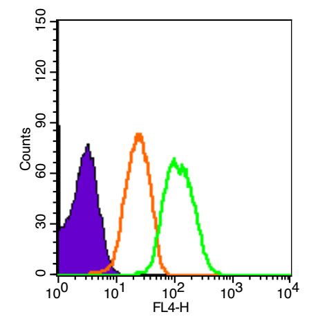
Fig1: Blank control (Black line): HUVEC(Black). Primary Antibody (green line): Rabbit Anti-XIAP antibody (ER1918-75) Dilution: 1μg /10^6 cells; Isotype Control Antibody (orange line): Rabbit IgG . Secondary Antibody (white blue line): Goat anti-rabbit IgG-PE Dilution: 1μg /test. Protocol The cells were fixed with 4% PFA (10min at room temperature)and then permeabilized with 90% ice-cold methanol for 20 min at room temperature. The cells were then incubated in 5%BSA to block non-specific protein-protein interactions for 30 min at room temperature .Cells stained with Primary Antibody for 30 min at room temperature. The secondary antibody used for 40 min at room temperature. Acquisition of 20,000 events was performed.Application
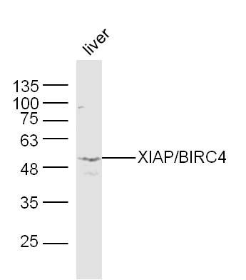
Fig2: Sample:Liver(Mouse) Lysate at 40 ug Primary: Anti-XIAP/BIRC4(ER1918-75) at 1/300 dilution Secondary: IRDye800CW Goat Anti-Rabbit IgG at 1/20000 dilution Predicted band size: 55 kD Observed band size: 53 kDApplication
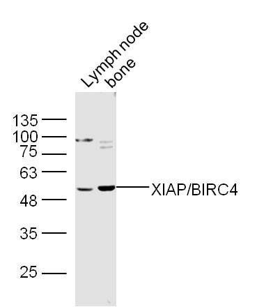
Fig3: Sample: Lymph node(Mouse) Lysate at 40 ug Bone(Mouse) Lysate at 40 ug Primary: Anti-XIAP/BIRC4(ER1918-75) at 1/300 dilution Secondary: IRDye800CW Goat Anti-Rabbit IgG at 1/20000 dilution Predicted band size: 55 kD Observed band size: 53 kDApplication
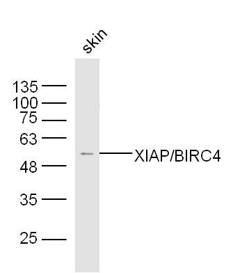
Fig4: Sample:Skin(Mouse) Lysate at 40 ug Primary: Anti-XIAP/BIRC4(ER1918-75) at 1/300 dilution Secondary: IRDye800CW Goat Anti-Rabbit IgG at 1/20000 dilution Predicted band size: 55 kD Observed band size: 53 kDApplication
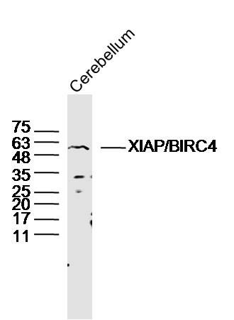
Fig5: Sample: Cerebellum (Mouse) Lysate at 40 ug Primary: Anti-XIAP/BIRC4 (ER1918-75) at 1/300 dilution Secondary: IRDye800CW Goat Anti-Rabbit IgG at 1/20000 dilution Predicted band size: 55 kD Observed band size: 55 kD| Product Name | XIAP/BIRC4 |
|---|---|
| Antibody Type | Primary Antibodies |
| Antigen Alias | X-linked Inhibitor of Apoptosis Protein RIAP-3 Baculoviral IAP repeat-containing protein 4 E3 ubiquitin-protein ligase XIAP Inhibitor of apoptosis protein 3 X-linked IAP IAP-like protein HILP BIRC4 HILP XLP2 ILP1 Xiap MIHA hILP IAP3 API3 XIAP XIAP_HUMAN ILP hILP IAP-3 hIAP-3 hIAP3 X-linked inhibitor of apoptosis protein. |
| Product description | Apoptotic suppressor. Has E3 ubiquitin-protein ligase activity. Mediates the proteasomal degradation of target proteins, such as caspase-3, SMAC or AIFM1. Inhibitor of caspase-3, -7 and -9. Mediates activation of MAP3K7/TAK1, leading to the activation of NF-kappa-B. |
| Immunogen | KLH conjugated synthetic peptide derived from human XIAP 201-330/496 |
| Clonality | Polyclonal |
|---|---|
| Isotype | IgG |
| Host Species | Rabbit |
| Tested Applications | WBFC |
| WB:1:500-2000: FC:3ug/Test: |
|
| Species Reactivity | HumanMouseRat |
| Concentration | 1mg/ml |
| Alternative Names | X-linked Inhibitor of Apoptosis Protein RIAP-3 Baculoviral IAP repeat-containing protein 4 E3 ubiquitin-protein ligase XIAP Inhibitor of apoptosis protein 3 X-linked IAP IAP-like protein HILP BIRC4 HILP XLP2 ILP1 Xiap MIHA hILP IAP3 API3 XIAP XIAP_HUMAN ILP hILP IAP-3 hIAP-3 hIAP3 X-linked inhibitor of apoptosis protein. |
|---|---|
| Molecular Weight(MW) | 55 KDa |
| Cellular Localization | Cytoplasm. |
| SwissProt ID | Q60989 |
|---|
Application
Fig1: Blank control (Black line): HUVEC(Black). Primary Antibody (green line): Rabbit Anti-XIAP antibody (ER1918-75) Dilution: 1μg /10^6 cells; Isotype Control Antibody (orange line): Rabbit IgG . Secondary Antibody (white blue line): Goat anti-rabbit IgG-PE Dilution: 1μg /test. Protocol The cells were fixed with 4% PFA (10min at room temperature)and then permeabilized with 90% ice-cold methanol for 20 min at room temperature. The cells were then incubated in 5%BSA to block non-specific protein-protein interactions for 30 min at room temperature .Cells stained with Primary Antibody for 30 min at room temperature. The secondary antibody used for 40 min at room temperature. Acquisition of 20,000 events was performed.
Application
Fig2: Sample:Liver(Mouse) Lysate at 40 ug Primary: Anti-XIAP/BIRC4(ER1918-75) at 1/300 dilution Secondary: IRDye800CW Goat Anti-Rabbit IgG at 1/20000 dilution Predicted band size: 55 kD Observed band size: 53 kD
Application
Fig3: Sample: Lymph node(Mouse) Lysate at 40 ug Bone(Mouse) Lysate at 40 ug Primary: Anti-XIAP/BIRC4(ER1918-75) at 1/300 dilution Secondary: IRDye800CW Goat Anti-Rabbit IgG at 1/20000 dilution Predicted band size: 55 kD Observed band size: 53 kD
Application
Fig4: Sample:Skin(Mouse) Lysate at 40 ug Primary: Anti-XIAP/BIRC4(ER1918-75) at 1/300 dilution Secondary: IRDye800CW Goat Anti-Rabbit IgG at 1/20000 dilution Predicted band size: 55 kD Observed band size: 53 kD
Application
Fig5: Sample: Cerebellum (Mouse) Lysate at 40 ug Primary: Anti-XIAP/BIRC4 (ER1918-75) at 1/300 dilution Secondary: IRDye800CW Goat Anti-Rabbit IgG at 1/20000 dilution Predicted band size: 55 kD Observed band size: 55 kD| Positive Control | Ubiquitous, except peripheral blood leukocytes. |
|---|---|
| Application Notes | WB:1:500-2000: FC:3ug/Test: |
| Form | Liquid |
|---|---|
| Storage Instructions | Store at -20℃ for one year. Avoid repeated freeze/thaw cycles. The lyophilized antibody is stable at room temperature for at least one month and for greater than a year when kept at -20℃. When reconstituted in sterile pH 7.4 0.01M PBS or diluent of antibody the antibody is stable for at least two weeks at 2-4℃. |
| Storage Buffer | 0.01M TBS(pH7.4) with 1% BSA, 0.03% Proclin300 and 50% Glycerol. |


