Application
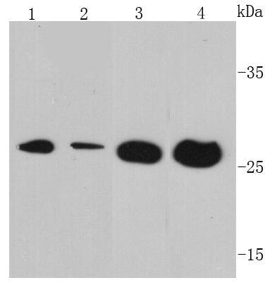
Fig1: Western blot analysis of VEGF on different cell lysates using anti- VEGF antibody at 1/500 dilution. Positive control: Lane 1: Jurkat Lane 2: Hela Lane 3: MCF-7 Lane 4: Human brainApplication
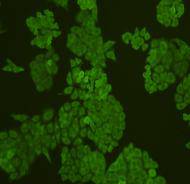
Fig2: ICC staining VEGF in HepG2 cells (green). Cells were fixed in paraformaldehyde, permeabilised with 0.25% Triton X100/PBS.Application
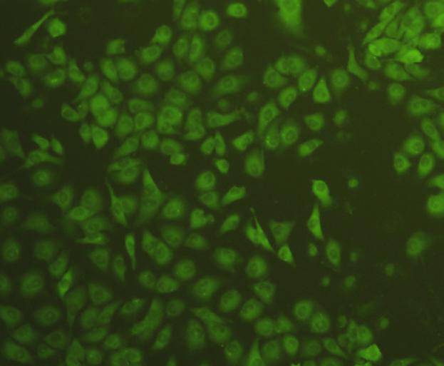
Fig3: ICC staining VEGF in HUVEC cells (green). Cells were fixed in paraformaldehyde, permeabilised with 0.25% Triton X100/PBS.Application
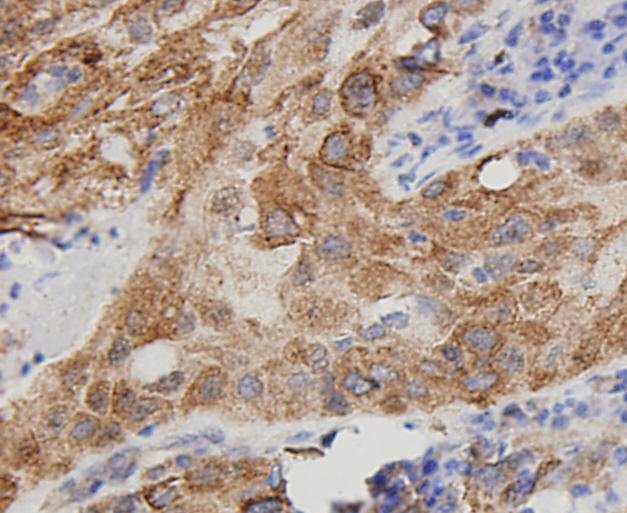
Fig4: Immunohistochemical analysis of paraffin-embedded human lung carcinoma tissue using anti-VEGF antibody. Counter stained with hematoxylin.Application
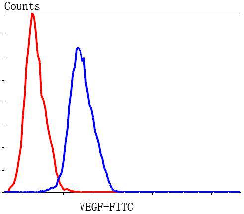
Fig5: Flow cytometric analysis of Hela cells with VEGF antibody at 1/100 dilution (blue) compared with an unlabelled control (cells without incubation with primary antibody; red). Goat anti rabbit IgG (FITC) was used as the secondary antibody.| Product Name | VEGF |
|---|---|
| Antibody Type | Primary Antibodies |
| Antigen Alias | Folliculostellate cell-derived growth factor antibody Glioma-derived endothelial cell mitogen antibody MGC70609 antibody MVCD1 antibody Vascular endothelial growth factor A antibody vascular endothelial growth factor A121 antibody vascular endothelial growth factor A165 antibody vascular endothelial growth factor antibody Vascular permeability factor antibody VEGF A antibody Vegf antibody VEGF-A antibody VEGF120 antibody Vegfa antibody VEGFA_HUMAN antibody VPF antibody |
| Product description | Vascular endothelial growth factor A (VEGFA) is a dimeric glycoprotein that plays a significant role in neurons and is considered to be the main, dominant inducer to the growth of blood vessels. VEGFA is essential for adults during organ remodeling and diseases that involve blood vessels, for example, in wound healing, tumor angiogenesis, diabetic retinopathy, and age-related muscular degeneration. |
| Immunogen | peptide |
| Clonality | Polyclonal |
|---|---|
| Isotype | IgG |
| Host Species | Rabbit |
| Tested Applications | WBICCIHCFC |
| WB:1:500-1:1,000 ICC:1:200 IHC:1:200 FC:1:100-1:200 |
|
| Species Reactivity | Human |
| Concentration | 1mg/ml |
| Alternative Names | Folliculostellate cell-derived growth factor antibody Glioma-derived endothelial cell mitogen antibody MGC70609 antibody MVCD1 antibody Vascular endothelial growth factor A antibody vascular endothelial growth factor A121 antibody vascular endothelial growth factor A165 antibody vascular endothelial growth factor antibody Vascular permeability factor antibody VEGF A antibody Vegf antibody VEGF-A antibody VEGF120 antibody Vegfa antibody VEGFA_HUMAN antibody VPF antibody |
|---|---|
| Molecular Weight(MW) | 27 kDa |
| Cellular Localization | Secreted. |
| SwissProt ID | P15692 |
|---|
Application
Fig1: Western blot analysis of VEGF on different cell lysates using anti- VEGF antibody at 1/500 dilution. Positive control: Lane 1: Jurkat Lane 2: Hela Lane 3: MCF-7 Lane 4: Human brain
Application
Fig2: ICC staining VEGF in HepG2 cells (green). Cells were fixed in paraformaldehyde, permeabilised with 0.25% Triton X100/PBS.
Application
Fig3: ICC staining VEGF in HUVEC cells (green). Cells were fixed in paraformaldehyde, permeabilised with 0.25% Triton X100/PBS.
Application
Fig4: Immunohistochemical analysis of paraffin-embedded human lung carcinoma tissue using anti-VEGF antibody. Counter stained with hematoxylin.
Application
Fig5: Flow cytometric analysis of Hela cells with VEGF antibody at 1/100 dilution (blue) compared with an unlabelled control (cells without incubation with primary antibody; red). Goat anti rabbit IgG (FITC) was used as the secondary antibody.| Positive Control | MCF-7, Hela, Jurkat, HUVEC, human brain tissue, human lung carcinoma tissue, HepG2. |
|---|---|
| Application Notes | WB:1:500-1:1,000 ICC:1:200 IHC:1:200 FC:1:100-1:200 |
| Form | Liquid |
|---|---|
| Storage Instructions | Store at +4℃ after thawing. Aliquot store at -20℃ or -80℃. Avoid repeated freeze / thaw cycles. |
| Storage Buffer | 1*TBS (pH7.4), 1%BSA, 40%Glycerol. Preservative: 0.05% Sodium Azide. |


