Application
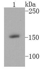
Fig1: Western blot analysis of MSH6 on A431 cell lysates using anti-MSH6 antibody at 1/1000 dilution.Application
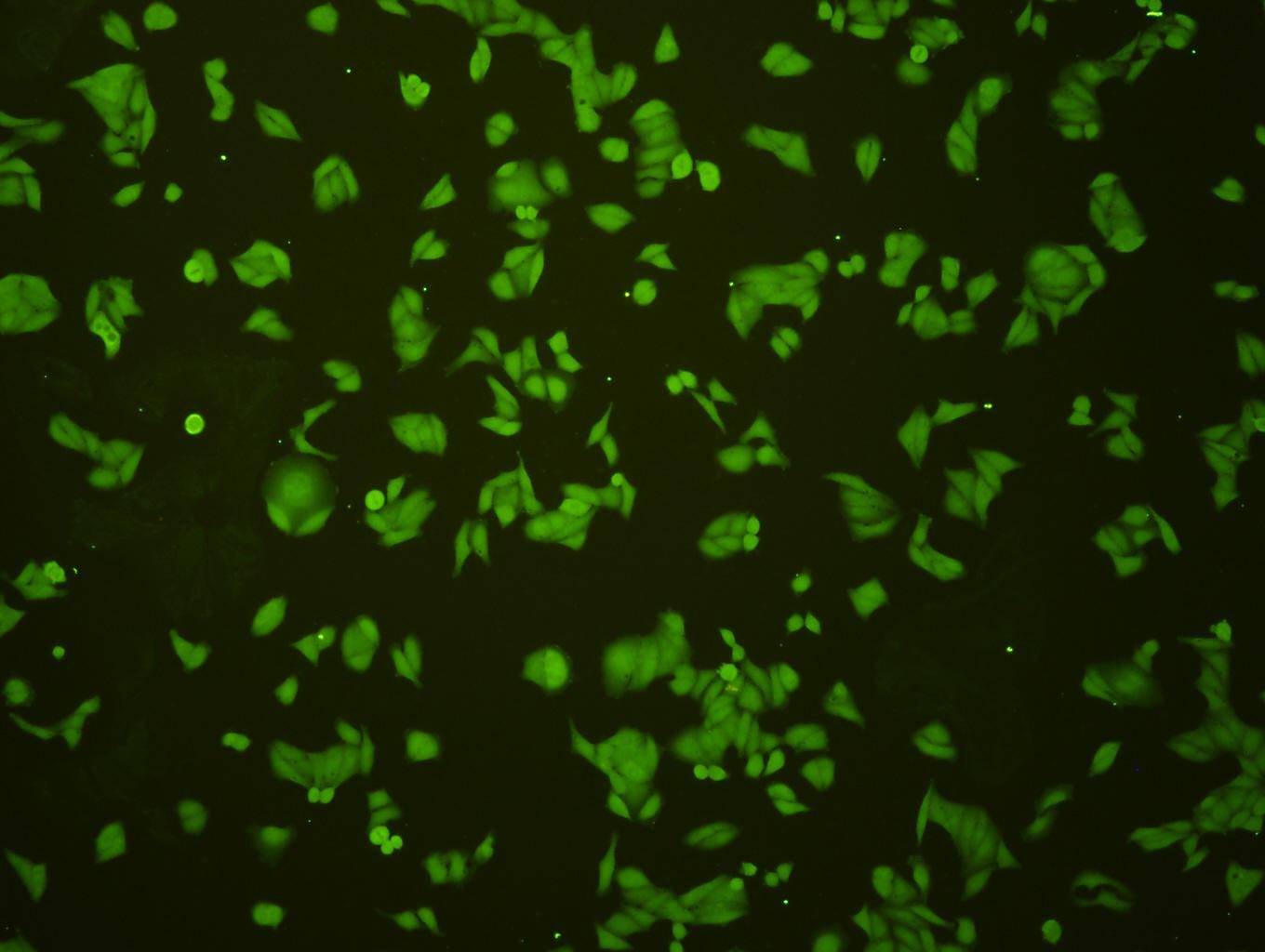
Fig2: ICC staining MSH6 in Hela cells (green). Cells were fixed in paraformaldehyde, permeabilised with 0.25% Triton X100/PBS.Application
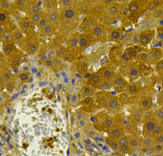
Fig3: Immunohistochemical analysis of paraffin-embedded rat liver tissue using anti-MSH6 antibody. Counter stained with hematoxylin.Application
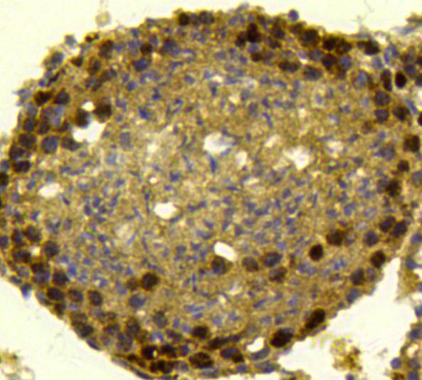
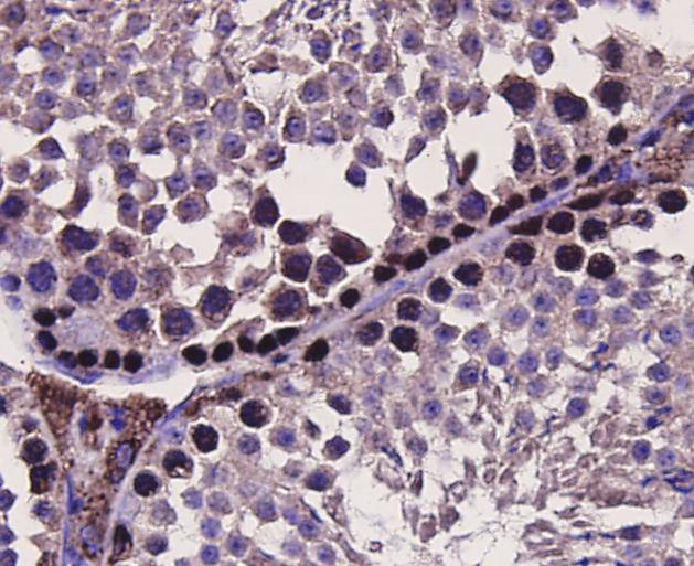
Fig4: Immunohistochemical analysis of paraffin-embedded rat testicle tissue using anti-MSH6 antibody. Counter stained with hematoxylin.Application
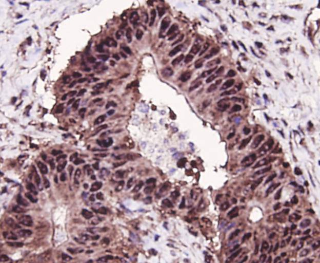
Fig5: Immunohistochemical analysis of paraffin-embedded human colon cancer tissue using anti-MSH6 antibody. Counter stained with hematoxylin.Application
Fig6: Immunohistochemical analysis of paraffin-embedded mouse testicle tissue using anti-MSH6 antibody. Counter stained with hematoxylin.| Product Name | MSH6 |
|---|---|
| Antibody Type | Primary Antibodies |
| Antigen Alias | DNA mismatch repair protein Msh6 antibody G/T mismatch binding protein antibody G/T mismatch-binding protein antibody GTBP antibody GTMBP antibody hMSH6 antibody HNPCC 5 antibody HNPCC5 antibody HSAP antibody MSH 6 antibody MSH6 antibody MSH6_HUMAN antibody mutS (E. coli) homolog 6 antibody MutS alpha 160 kDa subunit antibody MutS homolog 6 (E. coli) antibody mutS homolog 6 antibody MutS-alpha 160 kDa subunit antibody p160 antibody Sperm associated protein antibody |
| Product description | The DNA mismatch repair system (MMR) repairs post-replication DNA, inhibits recombination between nonidentical DNA sequences, and induces both checkpoint and apoptotic responses following certain types of DNA damage. MSH6 heterodimerizes with MSH2 to form MutS alpha, which binds to DNA mismatches thereby initiating DNA repair. Mutations in MSH6 and other MMR proteins have been found in a large proportion of hereditary nonpolyposis colorectal cancer (Lynch Syndrome), the most common form of inherited colorectal cancer in the Western world. |
| Immunogen | peptide |
| Clonality | Polyclonal |
|---|---|
| Isotype | IgG |
| Host Species | Rabbit |
| Tested Applications | WBICCIHCFC |
| WB:1:1,000 ICC:1:200 IHC:1:200 FC:1:100-1:200 |
|
| Species Reactivity | HumanMouseRat |
| Concentration | 1mg/ml |
| Alternative Names | DNA mismatch repair protein Msh6 antibody G/T mismatch binding protein antibody G/T mismatch-binding protein antibody GTBP antibody GTMBP antibody hMSH6 antibody HNPCC 5 antibody HNPCC5 antibody HSAP antibody MSH 6 antibody MSH6 antibody MSH6_HUMAN antibody mutS (E. coli) homolog 6 antibody MutS alpha 160 kDa subunit antibody MutS homolog 6 (E. coli) antibody mutS homolog 6 antibody MutS-alpha 160 kDa subunit antibody p160 antibody Sperm associated protein antibody |
|---|---|
| Molecular Weight(MW) | 152 kDa |
| Cellular Localization | Nucleus, chromosome. |
| SwissProt ID | P52701 |
|---|
Application
Fig1: Western blot analysis of MSH6 on A431 cell lysates using anti-MSH6 antibody at 1/1000 dilution.
Application
Fig2: ICC staining MSH6 in Hela cells (green). Cells were fixed in paraformaldehyde, permeabilised with 0.25% Triton X100/PBS.
Application
Fig3: Immunohistochemical analysis of paraffin-embedded rat liver tissue using anti-MSH6 antibody. Counter stained with hematoxylin.
Application
Fig4: Immunohistochemical analysis of paraffin-embedded rat testicle tissue using anti-MSH6 antibody. Counter stained with hematoxylin.
Application
Fig5: Immunohistochemical analysis of paraffin-embedded human colon cancer tissue using anti-MSH6 antibody. Counter stained with hematoxylin.
Application
Fig6: Immunohistochemical analysis of paraffin-embedded mouse testicle tissue using anti-MSH6 antibody. Counter stained with hematoxylin.| Positive Control | Hela, A431, Lovo, mouse testicle tissue, rat liver tissue, rat testicle tissue, human colon cancer tissue. |
|---|---|
| Application Notes | WB:1:1,000 ICC:1:200 IHC:1:200 FC:1:100-1:200 |
| Form | Liquid |
|---|---|
| Storage Instructions | Store at +4℃ after thawing. Aliquot store at -20℃ or -80℃. Avoid repeated freeze / thaw cycles. |
| Storage Buffer | 1*TBS (pH7.4), 1%BSA, 40%Glycerol. Preservative: 0.05% Sodium Azide. |


