Application
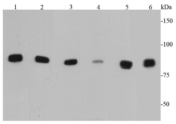
Fig1: Western blot analysis of IKK alpha on different cell lysates using anti-IKK alpha antibody at 1/500 dilution. Positive control: Lane 1: Hela Lane 2: HepG2 Lane 3: Raji Lane 4: NIH/3T3 Lane 5: PC12 Lane 6: JurkatApplication
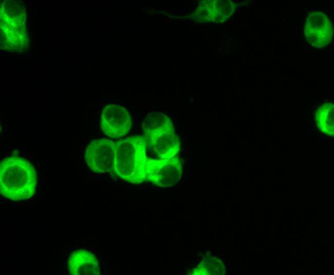
Fig2: ICC staining IKK alpha in N2A cells (green). Cells were fixed in paraformaldehyde, permeabilised with 0.25% Triton X100/PBS.Application
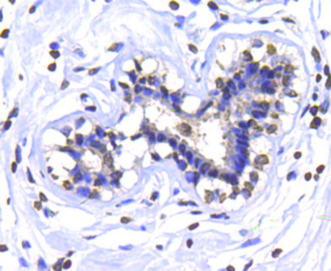
Fig3: Immunohistochemical analysis of paraffin-embedded human mammary gland tissue using anti-IKK alpha antibody. Counter stained with hematoxylin.Application
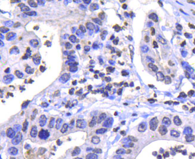
Fig4: Immunohistochemical analysis of paraffin-embedded human gastric carcinoma tissue using anti-IKK alpha antibody. Counter stained with hematoxylin.Application
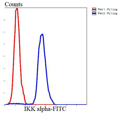
Fig5: Flow cytometric analysis of Hela cells with IKK alpha antibody at 1/100 dilution (blue) compared with an unlabelled control (cells without incubation with primary antibody; red). Goat anti rabbit IgG (FITC) was used as the secondary antibody.| Product Name | Ikk alpha |
|---|---|
| Antibody Type | Primary Antibodies |
| Antigen Alias | chuk antibody CHUK1 antibody Conserved Helix Loop Helix Ubiquitous Kinase antibody Conserved helix loop ubiquitous kinase antibody Conserved helix-loop-helix ubiquitous kinase antibody I Kappa B Kinase 1 antibody I Kappa B Kinase Alpha antibody I-kappa-B kinase 1 antibody I-kappa-B kinase alpha antibody IkappaB kinase antibody IkB kinase alpha subunit antibody IkBKA antibody IKK 1 antibody IKK A antibody IKK a kinase antibody IKK-A antibody IKK-alpha antibody IKK1 antibody IKKA antibody IKKA_HUMAN antibody Inhibitor Of Kappa Light Polypeptide Gene Enhancer In B Cells antibody Inhibitor Of Nuclear Factor Kappa B Kinase Alpha Subunit antibody Inhibitor of nuclear factor kappa-B kinase subunit alpha antibody NFKBIKA antibody Nuclear Factor Kappa B Inhibitor Kinase Alpha antibody Nuclear factor NF kappa B inhibitor kinase alpha antibody Nuclear factor NF-kappa-B inhibitor kinase alpha antibody Nuclear factor NFkappaB inhibitor kinase alpha antibody Nuclear Factor Of Kappa Light Chain Gene Enhancer In B Cells Inhibitor antibody TCF-16 antibody TCF16 antibody Transcription factor 16 antibody |
| Product description | Serine kinase that plays an essential role in the NF-kappa-B signaling pathway which is activated by multiple stimuli such as inflammatory cytokines, bacterial or viral products, DNA damages or other cellular stresses. It acts as part of the canonical IKK complex in the conventional pathway of NF-kappa-B activation and phosphorylates inhibitors of NF-kappa-B on serine residues. These modifications allow polyubiquitination of the inhibitors and subsequent degradation by the proteasome. In turn, free NF-kappa-B is translocated into the nucleus and activates the transcription of hundreds of genes involved in immune response, growth control, or protection against apoptosis. |
| Immunogen | peptide |
| Clonality | Polyclonal |
|---|---|
| Isotype | IgG |
| Host Species | Rabbit |
| Tested Applications | WBICCIHCFC |
| WB:1:500 ICC:1:200 IHC:1:200 FC:1:100-1:200 |
|
| Species Reactivity | HumanMouseRat |
| Concentration | 1mg/ml |
| Alternative Names | chuk antibody CHUK1 antibody Conserved Helix Loop Helix Ubiquitous Kinase antibody Conserved helix loop ubiquitous kinase antibody Conserved helix-loop-helix ubiquitous kinase antibody I Kappa B Kinase 1 antibody I Kappa B Kinase Alpha antibody I-kappa-B kinase 1 antibody I-kappa-B kinase alpha antibody IkappaB kinase antibody IkB kinase alpha subunit antibody IkBKA antibody IKK 1 antibody IKK A antibody IKK a kinase antibody IKK-A antibody IKK-alpha antibody IKK1 antibody IKKA antibody IKKA_HUMAN antibody Inhibitor Of Kappa Light Polypeptide Gene Enhancer In B Cells antibody Inhibitor Of Nuclear Factor Kappa B Kinase Alpha Subunit antibody Inhibitor of nuclear factor kappa-B kinase subunit alpha antibody NFKBIKA antibody Nuclear Factor Kappa B Inhibitor Kinase Alpha antibody Nuclear factor NF kappa B inhibitor kinase alpha antibody Nuclear factor NF-kappa-B inhibitor kinase alpha antibody Nuclear factor NFkappaB inhibitor kinase alpha antibody Nuclear Factor Of Kappa Light Chain Gene Enhancer In B Cells Inhibitor antibody TCF-16 antibody TCF16 antibody Transcription factor 16 antibody |
|---|---|
| Molecular Weight(MW) | 85 kDa |
| Cellular Localization | Cytoplasm, nucleus. |
| SwissProt ID | O15111 |
|---|
Application
Fig1: Western blot analysis of IKK alpha on different cell lysates using anti-IKK alpha antibody at 1/500 dilution. Positive control: Lane 1: Hela Lane 2: HepG2 Lane 3: Raji Lane 4: NIH/3T3 Lane 5: PC12 Lane 6: Jurkat
Application
Fig2: ICC staining IKK alpha in N2A cells (green). Cells were fixed in paraformaldehyde, permeabilised with 0.25% Triton X100/PBS.
Application
Fig3: Immunohistochemical analysis of paraffin-embedded human mammary gland tissue using anti-IKK alpha antibody. Counter stained with hematoxylin.
Application
Fig4: Immunohistochemical analysis of paraffin-embedded human gastric carcinoma tissue using anti-IKK alpha antibody. Counter stained with hematoxylin.
Application
Fig5: Flow cytometric analysis of Hela cells with IKK alpha antibody at 1/100 dilution (blue) compared with an unlabelled control (cells without incubation with primary antibody; red). Goat anti rabbit IgG (FITC) was used as the secondary antibody.| Positive Control | Hela, NIH/3T3, HepG2, Raji, PC12, N2A, Jurkat, human mammary gland tissue, human gastric carcinoma tissue. |
|---|---|
| Application Notes | WB:1:500 ICC:1:200 IHC:1:200 FC:1:100-1:200 |
| Form | Liquid |
|---|---|
| Storage Instructions | Store at +4℃ after thawing. Aliquot store at -20℃ or -80℃. Avoid repeated freeze / thaw cycles. |
| Storage Buffer | 1*TBS (pH7.4), 1%BSA, 40%Glycerol. Preservative: 0.05% Sodium Azide. |


