Application
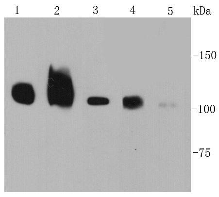
Fig1: Western blot analysis of CD29 on different cell lysates using anti-CD29 antibody at 1/1000 dilution. Positive control: Lane 1: Human liver Lane 2: Human kidney Lane 3: NIH/3T3 Lane 4: A172 Lane 5: HelaApplication
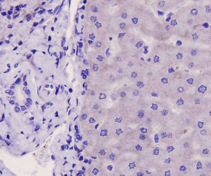
Fig2: Immunohistochemical analysis of paraffin-embedded human liver carcinoma tissue using anti-CD29 antibody. Counter stained with hematoxylin.Application
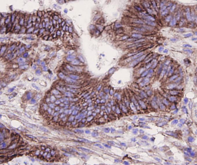
Fig3: Immunohistochemical analysis of paraffin-embedded human colon carcinoma tissue using anti-CD29 antibody. Counter stained with hematoxylin.Application
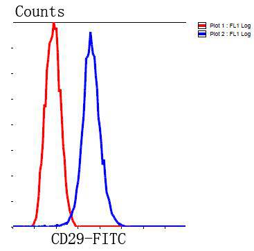
Fig4: Flow cytometric analysis of HepG2 cells with CD29 antibody at 1/100 dilution (blue) compared with an unlabelled control (cells without incubation with primary antibody; red). Goat anti rabbit IgG (FITC) was used as the secondary antibody.| Product Name | Integrin beta-1/CD29 |
|---|---|
| Antibody Type | Primary Antibodies |
| Antigen Alias | beta1 integrin antibody CD29 antibody Fibronectin receptor subunit beta antibody FNRB antibody Glycoprotein IIa antibody GP IIa antibody GPIIA antibody Integrin beta-1 antibody integrin VLA-4 beta subunit antibody Integrin, beta 1 (fibronectin receptor, beta polypeptide, antigen CD29 includes MDF2, MSK12) antibody ITB1_HUMAN antibody ITGB1 antibody MDF2 antibody MSK12 antibody OTTHUMP00000019420 antibody Very late activation protein, beta polypeptide antibody VLA BETA antibody VLA-4 subunit beta antibody VLA-BETA antibody VLAB antibody VLAbeta antibody |
| Product description | Integrins are transmembrane receptors that mediate the attachment between a cell and its surroundings, such as other cells or the extracellular matrix (ECM). Integrins are obligate heterodimers containing two distinct chains, called the α (alpha) and β (beta) subunits. The molecular mass of the integrin subunits can vary from 90?kDa to 160?kDa. Beta subunits have four cysteine-rich repeated sequences. Both α and β subunits bind several divalent cations. Integrins have two main functions: Attachment of the cell to the ECM and signal transduction from the ECM to the cell. However, they are also involved in a wide range of other biological activities, including immune patrolling, cell migration, and binding to cells by certain viruses, such as adenovirus, echovirus, hantavirus, and foot and mouth disease viruses. Research studies have implicated β1 integrin in various activities including embryonic development, blood vessel, skin, bone, and muscle formation, as well as tumor metastasis and angiogenesis. |
| Immunogen | peptide |
| Clonality | Polyclonal |
|---|---|
| Isotype | IgG |
| Host Species | Rabbit |
| Tested Applications | WBIHCFC |
| WB:1:500-1:1,000 FC:1:100 IHC:1:200 |
|
| Species Reactivity | HumanMouse |
| Concentration | 1mg/ml |
| Alternative Names | beta1 integrin antibody
CD29 antibody
Fibronectin receptor subunit beta antibody
FNRB antibody
Glycoprotein IIa antibody
GP IIa antibody
GPIIA antibody
Integrin beta-1 antibody
integrin VLA-4 beta subunit antibody
Integrin beta 1 (fibronectin receptor beta polypeptide antigen CD29 includes MDF2 MSK12) antibody ITB1_HUMAN antibody ITGB1 antibody MDF2 antibody MSK12 antibody OTTHUMP00000019420 antibody Very late activation protein beta polypeptide antibody VLA BETA antibody VLA-4 subunit beta antibody VLA-BETA antibody VLAB antibody VLAbeta antibody |
|---|---|
| Molecular Weight(MW) | 100 kDa |
| Cellular Localization | Cell membrane, cytoplasm |
| SwissProt ID | P05556 |
|---|
Application
Fig1: Western blot analysis of CD29 on different cell lysates using anti-CD29 antibody at 1/1000 dilution. Positive control: Lane 1: Human liver Lane 2: Human kidney Lane 3: NIH/3T3 Lane 4: A172 Lane 5: Hela
Application
Fig2: Immunohistochemical analysis of paraffin-embedded human liver carcinoma tissue using anti-CD29 antibody. Counter stained with hematoxylin.
Application
Fig3: Immunohistochemical analysis of paraffin-embedded human colon carcinoma tissue using anti-CD29 antibody. Counter stained with hematoxylin.
Application
Fig4: Flow cytometric analysis of HepG2 cells with CD29 antibody at 1/100 dilution (blue) compared with an unlabelled control (cells without incubation with primary antibody; red). Goat anti rabbit IgG (FITC) was used as the secondary antibody.| Positive Control | Human liver tissue, human kidney tissue, A172, NIH/3T3, Hela, human colonic carcinoma tissue, human liver cancer tissue, mouse kidney tissue, HepG2. |
|---|---|
| Application Notes | WB:1:500-1:1,000 FC:1:100 IHC:1:200 |
| Form | Liquid |
|---|---|
| Storage Instructions | Store at +4℃ after thawing. Aliquot store at -20℃ or -80℃. Avoid repeated freeze / thaw cycles. |
| Storage Buffer | 1*TBS (pH7.4), 1%BSA, 40%Glycerol. Preservative: 0.05% Sodium Azide. |


