Application
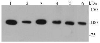
Fig1: Western blot analysis of CD133 on different cell lysates using anti-CD133 antibody at 1/1000 dilution.Application
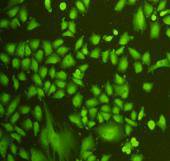
Fig2: ICC staining CD133 in A549 cells (green). Cells were fixed in paraformaldehyde, permeabilised with 0.25% Triton X100/PBS.Application
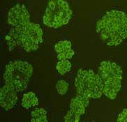
Fig3: ICC staining CD133 in HepG2 cells (green). Cells were fixed in paraformaldehyde, permeabilised with 0.25% Triton X100/PBS.Application
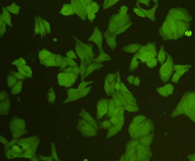
Fig4: ICC staining CD133 in Hela cells (green). Cells were fixed in paraformaldehyde, permeabilised with 0.25% Triton X100/PBS.Application
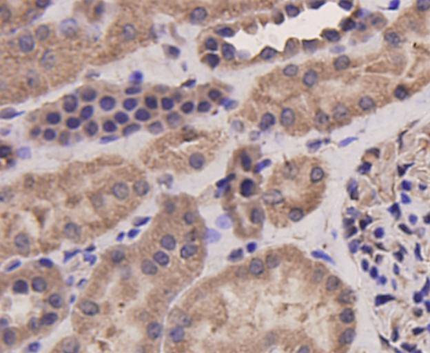
Fig5: Immunohistochemical analysis of paraffin-embedded human kidney tissue using anti-CD133 antibody. Counter stained with hematoxylin.Application
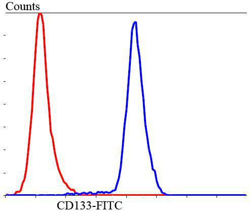
Fig6: Flow cytometric analysis of SHG-44 cells with CD133 antibody at 1/100 dilution (blue) compared with an unlabelled control (cells without incubation with primary antibody; red). Goat anti rabbit IgG (FITC) was used as the secondary antibody.Application
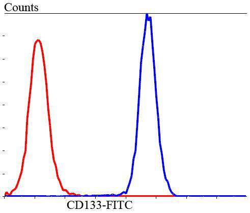
Fig7: Flow cytometric analysis of HUVEC cells with CD133 antibody at 1/100 dilution (blue) compared with an unlabelled control (cells without incubation with primary antibody; red). Goat anti rabbit IgG (FITC) was used as the secondary antibody.| Product Name | CD133 |
|---|---|
| Antibody Type | Primary Antibodies |
| Antigen Alias | AC133 antibody Antigen AC133 antibody CD133 antibody CORD12 antibody Hematopoietic stem cell antigen antibody hProminin antibody MCDR2 antibody MSTP061 antibody OTTHUMP00000217744 antibody OTTHUMP00000217745 antibody OTTHUMP00000217746 antibody PROM1 antibody PROM1_HUMAN antibody Prominin I antibody Prominin like 1 antibody Prominin like protein 1 precursor antibody Prominin mouse like 1 antibody Prominin-1 antibody Prominin-like protein 1 antibody Prominin1 antibody PROML1 antibody RP41 antibody STGD4 antibody |
| Product description | CD133 is a glycoprotein also known in humans and rodents as Prominin 1 (PROM1). It is the founding member of pentaspan transmembrane glycoproteins (5-transmembrane, 5-TM), which specifically localizes to cellular protrusions. CD133 is expressed in hematopoietic stem cells, endothelial progenitor cells, glioblastomas, neuronal and glial stem cells and some other cell types. CD133 is recognized as a stem cell marker for normal and cancerous tissues. |
| Immunogen | recombinant protein |
| Clonality | Polyclonal |
|---|---|
| Isotype | IgG |
| Host Species | Rabbit |
| Tested Applications | WBICCIHCFC |
| WB:1:1,000 ICC:1:200 IHC:1:200 FC:1:100-1:200 |
|
| Species Reactivity | Human |
| Concentration | 1mg/ml |
| Alternative Names | AC133 antibody Antigen AC133 antibody CD133 antibody CORD12 antibody Hematopoietic stem cell antigen antibody hProminin antibody MCDR2 antibody MSTP061 antibody OTTHUMP00000217744 antibody OTTHUMP00000217745 antibody OTTHUMP00000217746 antibody PROM1 antibody PROM1_HUMAN antibody Prominin I antibody Prominin like 1 antibody Prominin like protein 1 precursor antibody Prominin mouse like 1 antibody Prominin-1 antibody Prominin-like protein 1 antibody Prominin1 antibody PROML1 antibody RP41 antibody STGD4 antibody |
|---|---|
| Molecular Weight(MW) | 100 kDa |
| Cellular Localization | Cell membrane |
| SwissProt ID | O43490 |
|---|
Application
Fig1: Western blot analysis of CD133 on different cell lysates using anti-CD133 antibody at 1/1000 dilution.
Application
Fig2: ICC staining CD133 in A549 cells (green). Cells were fixed in paraformaldehyde, permeabilised with 0.25% Triton X100/PBS.
Application
Fig3: ICC staining CD133 in HepG2 cells (green). Cells were fixed in paraformaldehyde, permeabilised with 0.25% Triton X100/PBS.
Application
Fig4: ICC staining CD133 in Hela cells (green). Cells were fixed in paraformaldehyde, permeabilised with 0.25% Triton X100/PBS.
Application
Fig5: Immunohistochemical analysis of paraffin-embedded human kidney tissue using anti-CD133 antibody. Counter stained with hematoxylin.
Application
Fig6: Flow cytometric analysis of SHG-44 cells with CD133 antibody at 1/100 dilution (blue) compared with an unlabelled control (cells without incubation with primary antibody; red). Goat anti rabbit IgG (FITC) was used as the secondary antibody.
Application
Fig7: Flow cytometric analysis of HUVEC cells with CD133 antibody at 1/100 dilution (blue) compared with an unlabelled control (cells without incubation with primary antibody; red). Goat anti rabbit IgG (FITC) was used as the secondary antibody.| Positive Control | SW480, Hela, HepG2, HCT116, HT29, 293, HUVEC, SHG-44, human kidney tissue |
|---|---|
| Application Notes | WB:1:1,000 ICC:1:200 IHC:1:200 FC:1:100-1:200 |
| Form | Liquid |
|---|---|
| Storage Instructions | Store at +4℃ after thawing. Aliquot store at -20℃ or -80℃. Avoid repeated freeze / thaw cycles. |
| Storage Buffer | 1*TBS (pH7.4), 1%BSA, 40%Glycerol. Preservative: 0.05% Sodium Azide. |


