Application
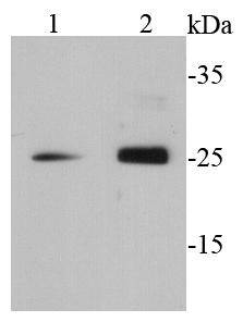
Fig1: Western blot analysis of GRB2 on different cell lysates using anti-GRB2 antibody at 1/500 dilution.Application
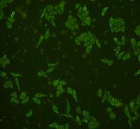
Fig2: ICC staining GRB2 in HepG2 cells (green). Cells were fixed in paraformaldehyde, permeabilised with 0.25% Triton X100/PBS.Application
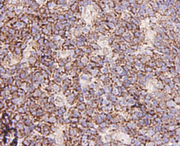
Fig3: Immunohistochemical analysis of paraffin-embedded human tonsil tissue using anti-GRB2 antibody. Counter stained with hematoxylin.Application
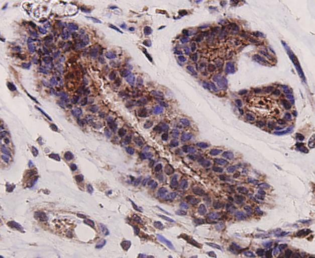
Fig4: Immunohistochemical analysis of paraffin-embedded human breast cancer tissue using anti-GRB2 antibody. Counter stained with hematoxylin.Application
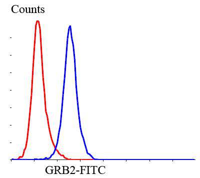
Fig5: Flow cytometric analysis of SHG-44 cells with GRB2 antibody at 1/100 dilution (blue) compared with an unlabelled control (cells without incubation with primary antibody; red). Goat anti rabbit IgG (FITC) was used as the secondary antibody.| Product Name | GRB2 |
|---|---|
| Antibody Type | Primary Antibodies |
| Antigen Alias | Abundant SRC homology antibody Adapter protein GRB2 antibody ASH antibody Ash protein antibody EGFRBP GRB2 antibody Epidermal growth factor receptor binding protein antibody Epidermal growth factor receptor binding protein GRB2 antibody GRB 2 antibody GRB2 adapter protein antibody Grb2 antibody GRB2_HUMAN antibody Grb3 3 antibody Growth factor receptor bound protein 2 antibody Growth factor receptor bound protein 3 antibody Growth factor receptor-bound protein 2 antibody HT027 antibody MST084 antibody MSTP084 antibody NCKAP2 antibody OTTHUMP00000166096 antibody OTTHUMP00000166097 antibody OTTHUMP00000166098 antibody Protein ash antibody SEM5 antibody SH2/SH3 adapter GRB2 antibody |
| Product description | Grb2 is widely expressed and is essential for multiple cellular functions. Inhibition of Grb2 function impairs developmental processes in various organisms and blocks transformation and proliferation of various cell types, and so it is not surprising that a targeted gene disruption of Grb2 in mouse is lethal at an early embryonic stage. Grb2 is best known for its ability to link the epidermal growth factor receptor tyrosine kinase to the activation of Ras and its downstream kinases, ERK1,2. Grb2 is composed of an SH2 domain flanked on each side by an SH3 domain. Grb2 has two closely related proteins with similar domain organizations, Gads and Grap. Gads and Grap are expressed specifically in hematopoietic cells and function in the coordination of tyrosine kinase mediated signal transduction. The SH2 domain of Grb2 binds to phosphorylated tyrosine-containing peptides on receptors or scaffold proteins with a preference for pY-X-N-X, where X is generally a hydrophobic residue such as valine. |
| Immunogen | peptide |
| Clonality | Polyclonal |
|---|---|
| Isotype | IgG |
| Host Species | Rabbit |
| Tested Applications | WBICCIHCFC |
| WB:1:500 ICC:1:200 IHC:1:200 FC:1:100-1:200 |
|
| Species Reactivity | HumanMouseRat |
| Concentration | 1mg/ml |
| Alternative Names | Abundant SRC homology antibody Adapter protein GRB2 antibody ASH antibody Ash protein antibody EGFRBP GRB2 antibody Epidermal growth factor receptor binding protein antibody Epidermal growth factor receptor binding protein GRB2 antibody GRB 2 antibody GRB2 adapter protein antibody Grb2 antibody GRB2_HUMAN antibody Grb3 3 antibody Growth factor receptor bound protein 2 antibody Growth factor receptor bound protein 3 antibody Growth factor receptor-bound protein 2 antibody HT027 antibody MST084 antibody MSTP084 antibody NCKAP2 antibody OTTHUMP00000166096 antibody OTTHUMP00000166097 antibody OTTHUMP00000166098 antibody Protein ash antibody SEM5 antibody SH2/SH3 adapter GRB2 antibody |
|---|---|
| Molecular Weight(MW) | 25 kDa |
| Cellular Localization | Cytoplasm, nucleus. |
| SwissProt ID | P62993 |
|---|
Application
Fig1: Western blot analysis of GRB2 on different cell lysates using anti-GRB2 antibody at 1/500 dilution.
Application
Fig2: ICC staining GRB2 in HepG2 cells (green). Cells were fixed in paraformaldehyde, permeabilised with 0.25% Triton X100/PBS.
Application
Fig3: Immunohistochemical analysis of paraffin-embedded human tonsil tissue using anti-GRB2 antibody. Counter stained with hematoxylin.
Application
Fig4: Immunohistochemical analysis of paraffin-embedded human breast cancer tissue using anti-GRB2 antibody. Counter stained with hematoxylin.
Application
Fig5: Flow cytometric analysis of SHG-44 cells with GRB2 antibody at 1/100 dilution (blue) compared with an unlabelled control (cells without incubation with primary antibody; red). Goat anti rabbit IgG (FITC) was used as the secondary antibody.| Positive Control | F9, HepG2, SHG-44, human breast cancer tissue, human tonsil tissue. |
|---|---|
| Application Notes | WB:1:500 ICC:1:200 IHC:1:200 FC:1:100-1:200 |
| Form | Liquid |
|---|---|
| Storage Instructions | Store at +4℃ after thawing. Aliquot store at -20℃ or -80℃. Avoid repeated freeze / thaw cycles. |
| Storage Buffer | 1*TBS (pH7.4), 1%BSA, 40%Glycerol. Preservative: 0.05% Sodium Azide. |


