Application
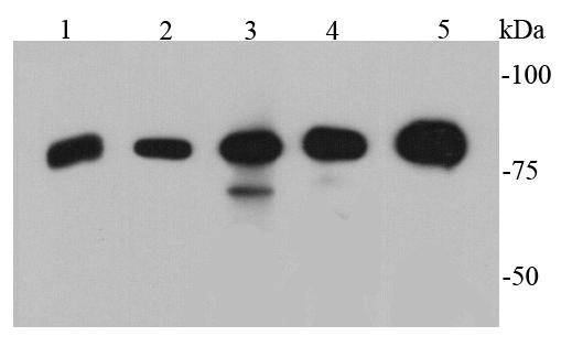
Fig1: Western blot analysis of p70 S6 Kinase beta on different cell lysates using anti-p70 S6 Kinase beta antibody at 1/500 dilution. Positive control: Lane 1: PC12 Lane 2: NIH/3T3 Lane 3: Jurkat Lane 4: K562 Lane 5: HelaApplication
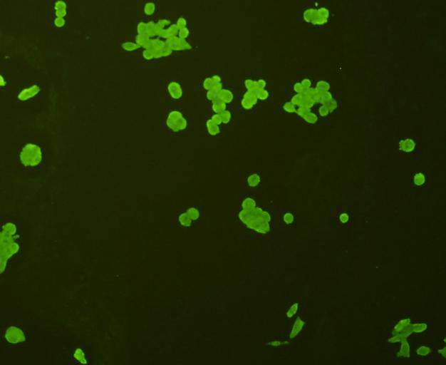
Fig2: ICC staining p70 S6 Kinase beta in Lovo cells (green). Cells were fixed in paraformaldehyde, permeabilised with 0.25% Triton X100/PBS.Application
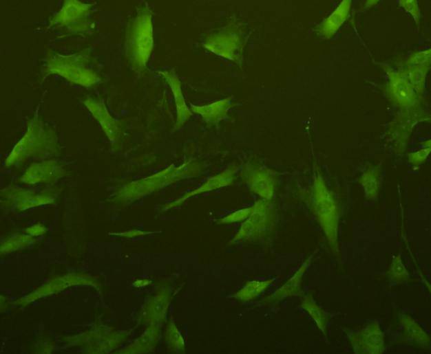
Fig3: ICC staining p70 S6 Kinase beta in NIH/3T3 cells (green). Cells were fixed in paraformaldehyde, permeabilised with 0.25% Triton X100/PBS.Application
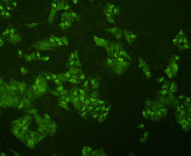
Fig4: ICC staining p70 S6 Kinase beta in Hela cells (green). Cells were fixed in paraformaldehyde, permeabilised with 0.25% Triton X100/PBS.Application
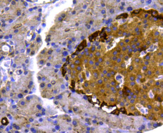
Fig5: Immunohistochemical analysis of paraffin-embedded rat pancreas tissue using anti- p70 S6 Kinase beta antibody. Counter stained with hematoxylin.Application
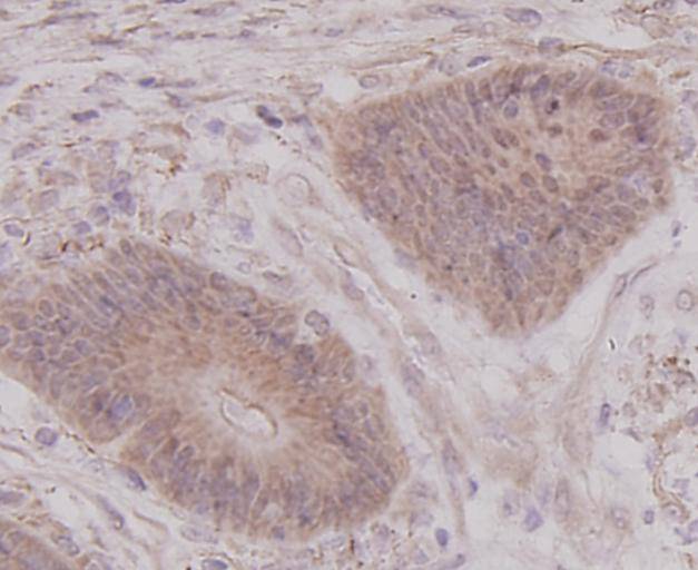
Fig6: Immunohistochemical analysis of paraffin-embedded human colon cancer tissue using anti- p70 S6 Kinase beta antibody. Counter stained with hematoxylin.Application
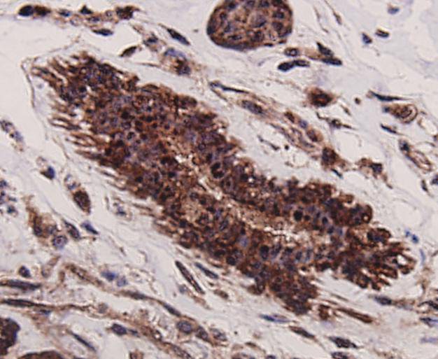
Fig7: Immunohistochemical analysis of paraffin-embedded human breast cancer tissue using anti- p70 S6 Kinase beta antibody. Counter stained with hematoxylin.Application
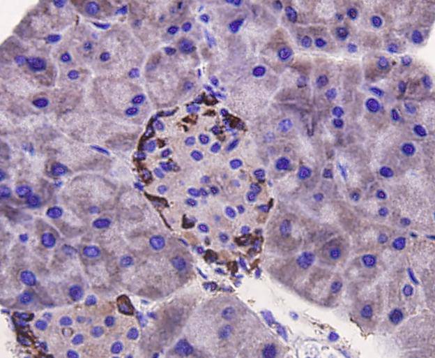
Fig8: Immunohistochemical analysis of paraffin-embedded mouse pancreas tissue using anti- p70 S6 Kinase beta antibody. Counter stained with hematoxylin.Application
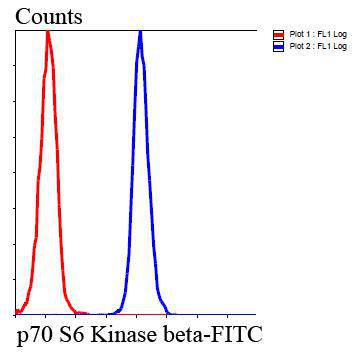
Fig9: Flow cytometric analysis of HepG2 cells with p70 S6 Kinase beta antibody at 1/100 dilution (blue) compared with an unlabelled control (cells without incubation with primary antibody; red). Goat anti rabbit IgG (FITC) was used as the secondary antibody.| Product Name | p70 S6 Kinase beta |
|---|---|
| Antibody Type | Primary Antibodies |
| Antigen Alias | 70 kDa ribosomal protein S6 kinase 2 antibody EC 2.7.11.1 antibody KS6B2_HUMAN antibody p70 beta antibody p70 ribosomal S6 kinase beta antibody p70 S6 kinase beta antibody p70 S6K-beta antibody p70 S6KB antibody p70 S6Kbeta antibody p70(S6K) beta antibody p70-beta antibody p70-S6K 2 antibody P70S6K2 antibody p70S6Kb antibody Ribosomal protein S6 kinase 70kDa, polypeptide 2 antibody Ribosomal protein S6 kinase beta 2 antibody Ribosomal protein S6 kinase beta-2 antibody Rps6kb2 antibody S6 kinase related kinase antibody S6 kinase-related kinase antibody S6K beta 2 antibody S6K beta antibody S6K-beta antibody S6K-beta-2 antibody S6K2 antibody Serine/threonine protein kinase 14 beta antibody Serine/threonine-protein kinase 14B antibody SRK antibody STK14B antibody |
| Product description | p70 S6 kinase is a mitogen activated Ser/Thr protein kinase that is required for cell growth and G1 cell cycle progression. p70 S6 kinase phosphorylates the S6 protein of the 40S ribosomal subunit and is involved in translational control of 5' oligopyrimidine tract mRNAs. A second isoform, p85 S6 kinase, is derived from the same gene and is identical to p70 S6 kinase except for 23 extra residues at the amino terminus, which encode a nuclear localizing signal. Both isoforms lie on a mitogen activated signaling pathway downstream of phosphoinositide-3 kinase (PI-3K) and the target of rapamycin, FRAP/mTOR, a pathway distinct from the Ras/MAP kinase cascade. Ribosomal protein S6 kinase beta-2 is an enzyme that in humans is encoded by the RPS6KB2 gene. Phosphorylation of S6 leads to an increase in protein synthesis and cell proliferation. |
| Immunogen | peptide |
| Clonality | Polyclonal |
|---|---|
| Isotype | IgG |
| Host Species | Rabbit |
| Tested Applications | WBICCIHCFC |
| WB:1:500 ICC:1:200 IHC:1:200 FC:1:100-1:200 |
|
| Species Reactivity | HumanMouseRat |
| Concentration | 1mg/ml |
| Alternative Names | 70 kDa ribosomal protein S6 kinase 2 antibody
EC 2.7.11.1 antibody
KS6B2_HUMAN antibody
p70 beta antibody
p70 ribosomal S6 kinase beta antibody
p70 S6 kinase beta antibody
p70 S6K-beta antibody
p70 S6KB antibody
p70 S6Kbeta antibody
p70(S6K) beta antibody
p70-beta antibody
p70-S6K 2 antibody
P70S6K2 antibody
p70S6Kb antibody
Ribosomal protein S6 kinase 70kDa polypeptide 2 antibody Ribosomal protein S6 kinase beta 2 antibody Ribosomal protein S6 kinase beta-2 antibody Rps6kb2 antibody S6 kinase related kinase antibody S6 kinase-related kinase antibody S6K beta 2 antibody S6K beta antibody S6K-beta antibody S6K-beta-2 antibody S6K2 antibody Serine/threonine protein kinase 14 beta antibody Serine/threonine-protein kinase 14B antibody SRK antibody STK14B antibody |
|---|---|
| Molecular Weight(MW) | 70 kDa |
| Cellular Localization | Cytoplasm, Nucleus |
| SwissProt ID | Q9UBS0 |
|---|
Application
Fig1: Western blot analysis of p70 S6 Kinase beta on different cell lysates using anti-p70 S6 Kinase beta antibody at 1/500 dilution. Positive control: Lane 1: PC12 Lane 2: NIH/3T3 Lane 3: Jurkat Lane 4: K562 Lane 5: Hela
Application
Fig2: ICC staining p70 S6 Kinase beta in Lovo cells (green). Cells were fixed in paraformaldehyde, permeabilised with 0.25% Triton X100/PBS.
Application
Fig3: ICC staining p70 S6 Kinase beta in NIH/3T3 cells (green). Cells were fixed in paraformaldehyde, permeabilised with 0.25% Triton X100/PBS.
Application
Fig4: ICC staining p70 S6 Kinase beta in Hela cells (green). Cells were fixed in paraformaldehyde, permeabilised with 0.25% Triton X100/PBS.
Application
Fig5: Immunohistochemical analysis of paraffin-embedded rat pancreas tissue using anti- p70 S6 Kinase beta antibody. Counter stained with hematoxylin.
Application
Fig6: Immunohistochemical analysis of paraffin-embedded human colon cancer tissue using anti- p70 S6 Kinase beta antibody. Counter stained with hematoxylin.
Application
Fig7: Immunohistochemical analysis of paraffin-embedded human breast cancer tissue using anti- p70 S6 Kinase beta antibody. Counter stained with hematoxylin.
Application
Fig8: Immunohistochemical analysis of paraffin-embedded mouse pancreas tissue using anti- p70 S6 Kinase beta antibody. Counter stained with hematoxylin.
Application
Fig9: Flow cytometric analysis of HepG2 cells with p70 S6 Kinase beta antibody at 1/100 dilution (blue) compared with an unlabelled control (cells without incubation with primary antibody; red). Goat anti rabbit IgG (FITC) was used as the secondary antibody.| Positive Control | PC12, Hela, NIH/3T3, Jurkat, HepG2, K562, Lovo, mouse pancreas tissue, rat pancreas tissue, human breast cancer tissue, human colon cancer tissue. |
|---|---|
| Application Notes | WB:1:500 ICC:1:200 IHC:1:200 FC:1:100-1:200 |
| Form | Liquid |
|---|---|
| Storage Instructions | Store at +4℃ after thawing. Aliquot store at -20℃ or -80℃. Avoid repeated freeze / thaw cycles. |
| Storage Buffer | 1*TBS (pH7.4), 1%BSA, 40%Glycerol. Preservative: 0.05% Sodium Azide. |


