Application
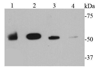
Fig1: Western blot analysis of MMP1 on different cell lysates using anti-MMP1 antibody at 1/1000 dilution. Positive control: Lane 1: SKBR3 Lane 2: Raji Lane 3: A431 Lane 4: A549Application
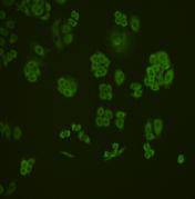
Fig2: ICC staining MMP1 in MCF-7 cells (green). Cells were fixed in paraformaldehyde, permeabilised with 0.25% Triton X100/PBS.Application
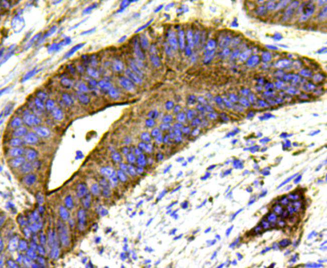
Fig3: Immunohistochemical analysis of paraffin-embedded human colon cancer tissue using anti-MMP1 antibody. Counter stained with hematoxylin.| Product Name | MMP1 |
|---|---|
| Antibody Type | Primary Antibodies |
| Antigen Alias | 27 kDa interstitial collagenase antibody CLG antibody CLGN antibody collagenase, fibroblast antibody collagenase, interstitial antibody Fibroblast collagenase antibody Interstitial collagenase antibody Matrix metallopeptidase 1 (interstitial collagenase) antibody Matrix metalloprotease 1 antibody Matrix Metalloproteinase 1 antibody Matrix metalloproteinase-1 antibody MMP 1 antibody MMP-1 antibody MMP1 antibody MMP1_HUMAN antibody OTTHUMP00000045866 antibody |
| Product description | MMP-1 has an archetypal structure consisting of a pre-domain, a pro-domain, a catalytic domain, a linker region and a hemopexin-like domain. Two main nomenclatures for the primary structure are currently in use, the original one from which the first amino-acid starts with the signalling peptide and a second one where the first amino-acid starts counting from the prodomain (proenzyme nomenclature). MMPs are involved in the breakdown of extracellular matrix in normal physiological processes, such as embryonic development, reproduction, and tissue remodeling, as well as in disease processes, such as arthritis and metastasis. Specifically, MMP-1 breaks down the interstitial collagens, types I, II, and III. Mechanical force may increase the expression of MMP1 in human periodontal ligament cells. |
| Immunogen | peptide |
| Clonality | Polyclonal |
|---|---|
| Isotype | IgG |
| Host Species | Rabbit |
| Tested Applications | WBICCIHC |
| WB:1:1,000 ICC:1:200 IHC:1:200 |
|
| Species Reactivity | Human |
| Concentration | 1mg/ml |
| Alternative Names | 27 kDa interstitial collagenase antibody
CLG antibody
CLGN antibody
collagenase fibroblast antibody collagenase interstitial antibody Fibroblast collagenase antibody Interstitial collagenase antibody Matrix metallopeptidase 1 (interstitial collagenase) antibody Matrix metalloprotease 1 antibody Matrix Metalloproteinase 1 antibody Matrix metalloproteinase-1 antibody MMP 1 antibody MMP-1 antibody MMP1 antibody MMP1_HUMAN antibody OTTHUMP00000045866 antibody |
|---|---|
| Molecular Weight(MW) | 54 kDa |
| Cellular Localization | Secreted. |
| SwissProt ID | P03956 |
|---|
Application
Fig1: Western blot analysis of MMP1 on different cell lysates using anti-MMP1 antibody at 1/1000 dilution. Positive control: Lane 1: SKBR3 Lane 2: Raji Lane 3: A431 Lane 4: A549
Application
Fig2: ICC staining MMP1 in MCF-7 cells (green). Cells were fixed in paraformaldehyde, permeabilised with 0.25% Triton X100/PBS.
Application
Fig3: Immunohistochemical analysis of paraffin-embedded human colon cancer tissue using anti-MMP1 antibody. Counter stained with hematoxylin.| Positive Control | A549, MCF-7, Raji, SKBR3, A431, human colon cancer tissue. |
|---|---|
| Application Notes | WB:1:1,000 ICC:1:200 IHC:1:200 |
| Form | Liquid |
|---|---|
| Storage Instructions | Store at +4℃ after thawing. Aliquot store at -20℃ or -80℃. Avoid repeated freeze / thaw cycles. |
| Storage Buffer | 1*TBS (pH7.4), 1%BSA, 40%Glycerol. Preservative: 0.05% Sodium Azide. |


