Application
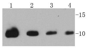
Fig1: Western blot analysis of Ubiquitin on different cell lysates using anti-Ubiquitin antibody at 1/1000 dilution. Positive control: Lane 1: 293T Lane 2: HepG2 Lane 3: Hela Lane 4: MCF-2Application
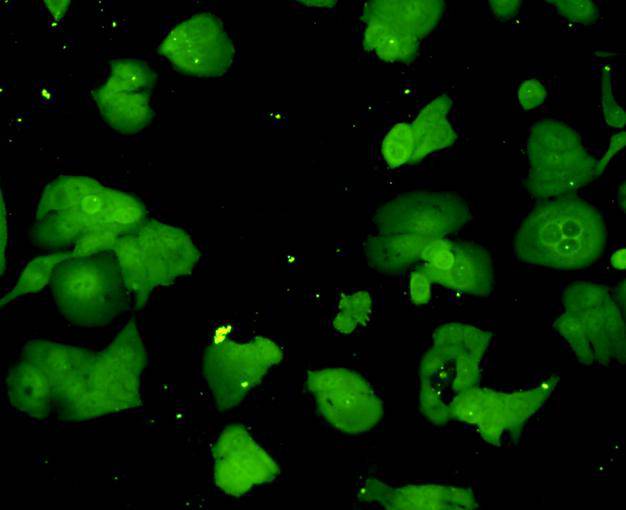
Fig2: ICC staining Ubiquitin in Hela cells (green). Cells were fixed in paraformaldehyde, permeabilised with 0.25% Triton X100/PBS.Application
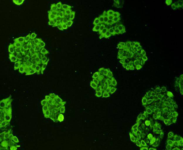
Fig3: ICC staining Ubiquitin in HepG2 cells (green). Cells were fixed in paraformaldehyde, permeabilised with 0.25% Triton X100/PBS.Application
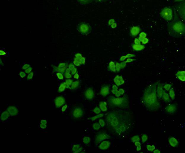
Fig4: ICC staining Ubiquitin in MCF-7 cells (green). Cells were fixed in paraformaldehyde, permeabilised with 0.25% Triton X100/PBS.Application
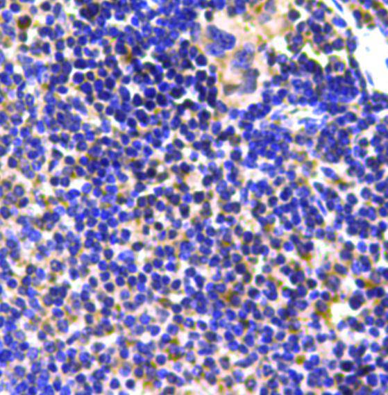
Fig5: Immunohistochemical analysis of paraffin-embedded mouse spleen tissue using anti-Ubiquitin antibody. Counter stained with hematoxylin.| Product Name | Ubiquitin |
|---|---|
| Antibody Type | Primary Antibodies |
| Antigen Alias | Epididymis secretory protein Li 50 antibody FLJ25987 antibody HEL S 50 antibody MGC8385 antibody Polyubiquitin B antibody RPS 27A antibody RPS27A antibody UBA 52 antibody UBA 80 antibody UBA52 antibody UBA80 antibody UBB antibody UBB_HUMAN antibody UBC antibody UBCEP 1 antibody UBCEP 2 antibody UBCEP1 antibody UBCEP2 antibody Ubiquitin antibody Ubiquitin B antibody |
| Product description | Ubiquitin is a conserved polypeptide unit that plays an important role in the ubiquitin-proteasome pathway. Ubiquitin exists either covalently attached to another protein, or free (unanchored). When covalently bound, it is conjugated to target proteins via an isopeptide bond either as a monomer (monoubiquitin), a polymer linked via different Lys residues of the ubiquitin (polyubiquitin chains) or a linear polymer linked via the initiator Met of the ubiquitin (linear polyubiquitin chains). Polyubiquitin chains, when attached to a target protein, have different functions depending on the Lys residue of the ubiquitin that is linked: Lys-6-linked may be involved in DNA repair; Lys-11-linked is involved in ERAD (endoplasmic reticulum-associated degradation) and in cell-cycle regulation; Lys-29-linked is involved in lysosomal degradation; Lys-33-linked is involved in kinase modification; Lys-48-linked is involved in protein degradation via the proteasome; Lys-63-linked is involved in endocytosis, DNA-damage responses as well as in signaling processes leading to activation of the transcription factor NF-kappa-B. |
| Immunogen | peptide |
| Clonality | Polyclonal |
|---|---|
| Isotype | IgG |
| Host Species | Rabbit |
| Tested Applications | WBICCIHC |
| WB:1:500-1:1,000 ICC:1:200 IHC:1:200 |
|
| Species Reactivity | HumanMouseRat |
| Concentration | 1mg/ml |
| Alternative Names | Epididymis secretory protein Li 50 antibody FLJ25987 antibody HEL S 50 antibody MGC8385 antibody Polyubiquitin B antibody RPS 27A antibody RPS27A antibody UBA 52 antibody UBA 80 antibody UBA52 antibody UBA80 antibody UBB antibody UBB_HUMAN antibody UBC antibody UBCEP 1 antibody UBCEP 2 antibody UBCEP1 antibody UBCEP2 antibody Ubiquitin antibody Ubiquitin B antibody |
|---|---|
| Molecular Weight(MW) | 10 kDa |
| Cellular Localization | Cytoplasm, nucleus |
| SwissProt ID | P0CG48 |
|---|
Application
Fig1: Western blot analysis of Ubiquitin on different cell lysates using anti-Ubiquitin antibody at 1/1000 dilution. Positive control: Lane 1: 293T Lane 2: HepG2 Lane 3: Hela Lane 4: MCF-2
Application
Fig2: ICC staining Ubiquitin in Hela cells (green). Cells were fixed in paraformaldehyde, permeabilised with 0.25% Triton X100/PBS.
Application
Fig3: ICC staining Ubiquitin in HepG2 cells (green). Cells were fixed in paraformaldehyde, permeabilised with 0.25% Triton X100/PBS.
Application
Fig4: ICC staining Ubiquitin in MCF-7 cells (green). Cells were fixed in paraformaldehyde, permeabilised with 0.25% Triton X100/PBS.
Application
Fig5: Immunohistochemical analysis of paraffin-embedded mouse spleen tissue using anti-Ubiquitin antibody. Counter stained with hematoxylin.| Positive Control | 293T, HepG2, Hela, MCF-7, human tonsil tissue,mouse spleen tissue. |
|---|---|
| Application Notes | WB:1:500-1:1,000 ICC:1:200 IHC:1:200 |
| Form | Liquid |
|---|---|
| Storage Instructions | Store at +4℃ after thawing. Aliquot store at -20℃ or -80℃. Avoid repeated freeze / thaw cycles. |
| Storage Buffer | 1*TBS (pH7.4), 1%BSA, 40%Glycerol. Preservative: 0.05% Sodium Azide. |


