Application
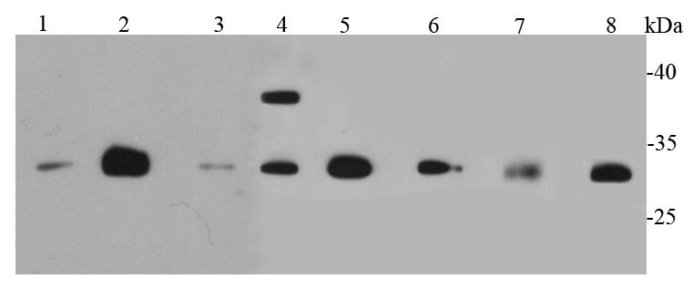
Fig1: Western blot analysis of CDk1 on different cell lysates using anti-CDk1 antibody at 1/500 dilution. Positive control: Lane 1: MCF-7 Lane 2: Jurkat Lane 3: PC12 Lane 4: HepG2 Lane 5: Hela Lane 6: NIH/3T3 Lane 7: Mouse liver Lane 8: SKBR3Application
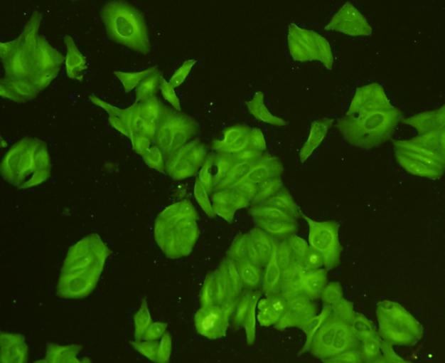
Fig2: ICC staining CDk1 in Hela cells (green). Cells were fixed in paraformaldehyde, permeabilised with 0.25% Triton X100/PBS.Application
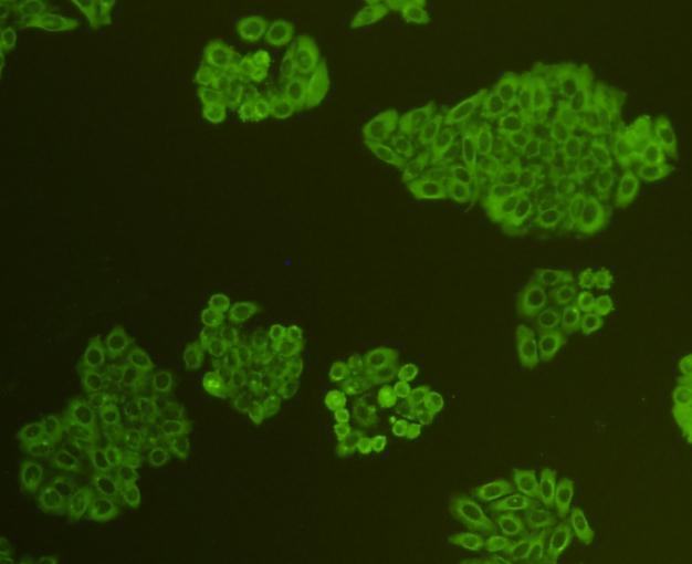
Fig3: ICC staining CDk1 in HepG2 cells (green). Cells were fixed in paraformaldehyde, permeabilised with 0.25% Triton X100/PBS.Application
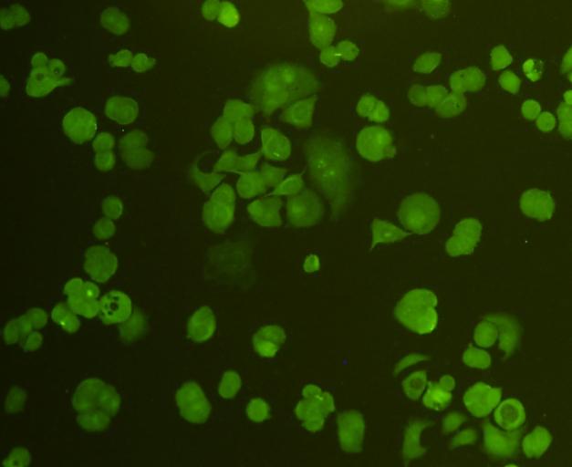
Fig4: ICC staining CDk1 in MCF-7 cells (green). Cells were fixed in paraformaldehyde, permeabilised with 0.25% Triton X100/PBS.Application
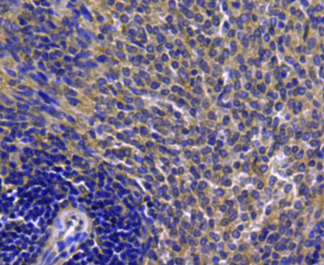
Fig5: Immunohistochemical analysis of paraffin-embedded rat spleen tissue using anti-CDk1 antibody. Counter stained with hematoxylin.Application
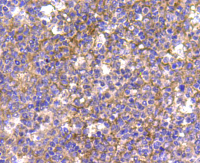
Fig6: Immunohistochemical analysis of paraffin-embedded human tonsil tissue using anti-CDk1 antibody. Counter stained with hematoxylinApplication
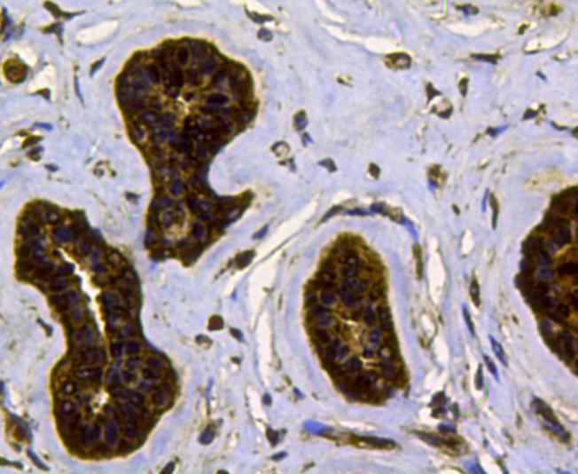
Fig7: Immunohistochemical analysis of paraffin-e.mbedded human breast cancer tissue using anti-CDk1 antibody. Counter stained with hematoxylinApplication
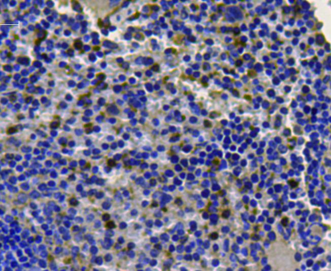
Fig8: Immunohistochemical analysis of paraffin-embedded mouse spleen tissue using anti-CDk1 antibody. Counter stained with hematoxylin.Application
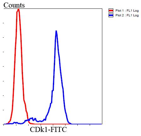
Fig9: Flow cytometric analysis of Hela cells with CDk1 antibody at 1/100 dilution (blue) compared with an unlabelled control (cells without incubation with primary antibody; red). Goat anti rabbit IgG (FITC) was used as the secondary antibody.| Product Name | CDk1 |
|---|---|
| Antibody Type | Primary Antibodies |
| Antigen Alias | Cdc 2 antibody Cdc2 antibody CDC28A antibody CDK 1 antibody CDK1 antibody CDK1_HUMAN antibody CDKN1 antibody CELL CYCLE CONTROLLER CDC2 antibody Cell division control protein 2 antibody Cell division control protein 2 homolog antibody Cell division cycle 2 G1 to S and G2 to M antibody Cell division protein kinase 1 antibody Cell Divsion Cycle 2 Protein antibody Cyclin Dependent Kinase 1 antibody Cyclin-dependent kinase 1 antibody DKFZp686L20222 antibody MGC111195 antibody p34 Cdk1 antibody p34 protein kinase antibody P34CDC2 antibody |
| Product description | Cdk1 is a small protein (approximately 34 kilodaltons), and is highly conserved. Cdk1 is comprised mostly by the bare protein kinase motif, which other protein kinases share. Cdk1, like other kinases, contains a cleft in which ATP fits. When bound to its cyclin partners, Cdk1 phosphorylation leads to cell cycle progression. Given its essential role in cell cycle progression, Cdk1 is highly regulated. Most obviously, Cdk1 is regulated by its binding with its cyclin partners. Cyclin binding alters access to the active site of Cdk1, allowing for Cdk1 activity; furthermore, cyclins impart specificity to Cdk1 activity. At least some cyclins contain a hydrophobic patch which may directly interact with substrates, conferring target specificity. Furthermore, cyclins can target Cdk1 to particular subcellular locations. |
| Immunogen | peptide |
| Clonality | Polyclonal |
|---|---|
| Isotype | IgG |
| Host Species | Rabbit |
| Tested Applications | WBICCIHCFC |
| WB:1:500 ICC:1:200 IHC:1:200 FC:1:100-1:200 |
|
| Species Reactivity | HumanMouseRat |
| Concentration | 1mg/ml |
| Alternative Names | Cdc 2 antibody Cdc2 antibody CDC28A antibody CDK 1 antibody CDK1 antibody CDK1_HUMAN antibody CDKN1 antibody CELL CYCLE CONTROLLER CDC2 antibody Cell division control protein 2 antibody Cell division control protein 2 homolog antibody Cell division cycle 2 G1 to S and G2 to M antibody Cell division protein kinase 1 antibody Cell Divsion Cycle 2 Protein antibody Cyclin Dependent Kinase 1 antibody Cyclin-dependent kinase 1 antibody DKFZp686L20222 antibody MGC111195 antibody p34 Cdk1 antibody p34 protein kinase antibody P34CDC2 antibody |
|---|---|
| Molecular Weight(MW) | 34 kDa |
| Cellular Localization | Cytoplasm, nucleus |
| SwissProt ID | P06493 |
|---|
Application
Fig1: Western blot analysis of CDk1 on different cell lysates using anti-CDk1 antibody at 1/500 dilution. Positive control: Lane 1: MCF-7 Lane 2: Jurkat Lane 3: PC12 Lane 4: HepG2 Lane 5: Hela Lane 6: NIH/3T3 Lane 7: Mouse liver Lane 8: SKBR3
Application
Fig2: ICC staining CDk1 in Hela cells (green). Cells were fixed in paraformaldehyde, permeabilised with 0.25% Triton X100/PBS.
Application
Fig3: ICC staining CDk1 in HepG2 cells (green). Cells were fixed in paraformaldehyde, permeabilised with 0.25% Triton X100/PBS.
Application
Fig4: ICC staining CDk1 in MCF-7 cells (green). Cells were fixed in paraformaldehyde, permeabilised with 0.25% Triton X100/PBS.
Application
Fig5: Immunohistochemical analysis of paraffin-embedded rat spleen tissue using anti-CDk1 antibody. Counter stained with hematoxylin.
Application
Fig6: Immunohistochemical analysis of paraffin-embedded human tonsil tissue using anti-CDk1 antibody. Counter stained with hematoxylin
Application
Fig7: Immunohistochemical analysis of paraffin-e.mbedded human breast cancer tissue using anti-CDk1 antibody. Counter stained with hematoxylin
Application
Fig8: Immunohistochemical analysis of paraffin-embedded mouse spleen tissue using anti-CDk1 antibody. Counter stained with hematoxylin.
Application
Fig9: Flow cytometric analysis of Hela cells with CDk1 antibody at 1/100 dilution (blue) compared with an unlabelled control (cells without incubation with primary antibody; red). Goat anti rabbit IgG (FITC) was used as the secondary antibody.| Positive Control | MCF-7, Jurkat, PC12, SKBR3, Hela, NIH/3T3, HepG2,rat liver tissue, mouse spleen tissue, human tonsil tissue, human breast cancer tissue, rat spleen tissue. |
|---|---|
| Application Notes | WB:1:500 ICC:1:200 IHC:1:200 FC:1:100-1:200 |
| Form | Liquid |
|---|---|
| Storage Instructions | Store at +4℃ after thawing. Aliquot store at -20℃ or -80℃. Avoid repeated freeze / thaw cycles. |
| Storage Buffer | 1*TBS (pH7.4), 1%BSA, 40%Glycerol. Preservative: 0.05% Sodium Azide. |


