Application
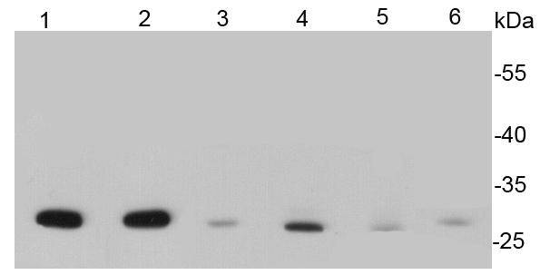
Fig1: Western blot analysis of Hsp27 on different cell lysates using anti- Hsp27 antibody at 1/500 dilution. Positive control: Lane 1: Hela Lane 2: Jurkat Lane 3: 293 Lane 4: Human kidney Lane 5: Mouse kidney Lane 6: Mouse heartApplication
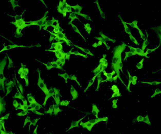
Fig2: ICC staining Hsp27 in A172 cells (green). Cells were fixed in paraformaldehyde, permeabilised with 0.25% Triton X100/PBS.Application
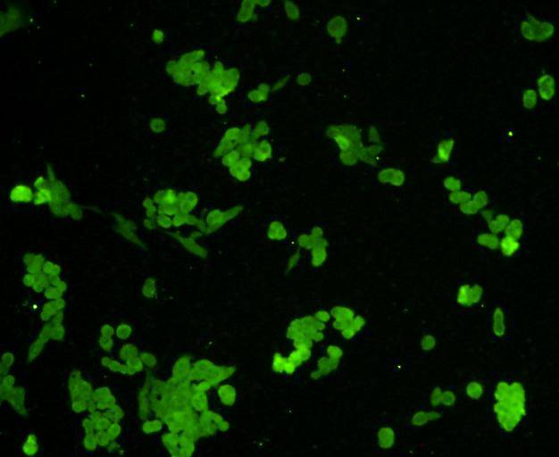
Fig3: ICC staining Hsp27 in Lovo cells (green). Cells were fixed in paraformaldehyde, permeabilised with 0.25% Triton X100/PBS.Application
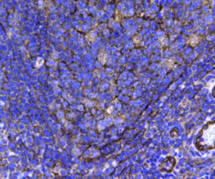
Fig4: Immunohistochemical analysis of paraffin-embedded human tonsil tissue using anti-Hsp27 antibody. Counter stained with hematoxylin.Application
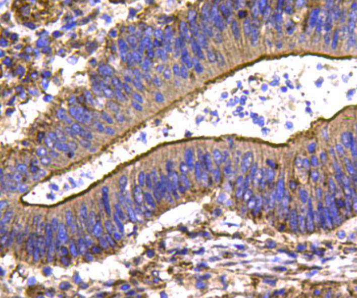
Fig5: Immunohistochemical analysis of paraffin-embedded human colon carcinoma tissue using anti-Hsp27 antibody. Counter stained with hematoxylin.| Product Name | HSP27 |
|---|---|
| Antibody Type | Primary Antibodies |
| Antigen Alias | Heat shock 27kDa protein antibody 28 kDa heat shock protein antibody CMT2F antibody DKFZp586P1322 antibody epididymis secretory protein Li 102 antibody Estrogen regulated 24 kDa protein antibody Estrogen-regulated 24 kDa protein antibody Heat shock 25kDa protein 1 antibody Heat shock 27 kDa protein antibody Heat shock 27kD protein 1 antibody Heat shock 27kDa protein 1 antibody Heat shock 28kDa protein 1 antibody Heat Shock Protein 27 antibody Heat shock protein beta 1 antibody Heat shock protein beta-1 antibody heat shock protein family B (small) member 1 antibody HEL-S-102 antibody HMN2B antibody HS.76067 antibody Hsp 25 antibody HSP 27 antibody Hsp 28 antibody Hsp B1 antibody Hsp25 antibody HSP27 antibody Hsp28 antibody HspB1 antibody HSPB1_HUMAN antibody SRP27 antibody Stress responsive protein 27 antibody Stress-responsive protein 27 antibody |
| Product description | Hsp27 is a chaperone of the sHsp (small heat shock protein) group among ubiquitin, α-crystallin, Hsp20 and others. The common functions of sHsps are chaperone activity, thermotolerance, inhibition of apoptosis, regulation of cell development, and cell differentiation. They also take part in signal transduction. Hsp27 appears in many cell types, especially all types of muscle cells. It is located mainly in the cytosol, but also in the perinuclear region, endoplasmatic reticulum, and nucleus. It is overexpressed during different stages of cell differentiation and development. This suggests an essential role for Hsp27 in the differentiation of tissues. Probably Hsp27 – among other chaperones – is involved in the process of cell differentiation. Changes of Hsp27 levels were observed in Ehrlich ascite cells, embryonic stem cells, normal B-cells, B-lymphoma cells, osteoblasts, keratinocytes, neurons etc. |
| Immunogen | peptide |
| Clonality | Polyclonal |
|---|---|
| Isotype | IgG |
| Host Species | Rabbit |
| Tested Applications | WBICCIHC |
| WB:1:500 ICC:1:100 IHC:1:200 |
|
| Species Reactivity | HumanMouseRat |
| Concentration | 1mg/ml |
| Alternative Names | Heat shock 27kDa protein antibody 28 kDa heat shock protein antibody CMT2F antibody DKFZp586P1322 antibody epididymis secretory protein Li 102 antibody Estrogen regulated 24 kDa protein antibody Estrogen-regulated 24 kDa protein antibody Heat shock 25kDa protein 1 antibody Heat shock 27 kDa protein antibody Heat shock 27kD protein 1 antibody Heat shock 27kDa protein 1 antibody Heat shock 28kDa protein 1 antibody Heat Shock Protein 27 antibody Heat shock protein beta 1 antibody Heat shock protein beta-1 antibody heat shock protein family B (small) member 1 antibody HEL-S-102 antibody HMN2B antibody HS.76067 antibody Hsp 25 antibody HSP 27 antibody Hsp 28 antibody Hsp B1 antibody Hsp25 antibody HSP27 antibody Hsp28 antibody HspB1 antibody HSPB1_HUMAN antibody SRP27 antibody Stress responsive protein 27 antibody Stress-responsive protein 27 antibody |
|---|---|
| Molecular Weight(MW) | 27 kDa |
| Cellular Localization | Cytoplasm, nucleus |
| SwissProt ID | P04792 |
|---|
Application
Fig1: Western blot analysis of Hsp27 on different cell lysates using anti- Hsp27 antibody at 1/500 dilution. Positive control: Lane 1: Hela Lane 2: Jurkat Lane 3: 293 Lane 4: Human kidney Lane 5: Mouse kidney Lane 6: Mouse heart
Application
Fig2: ICC staining Hsp27 in A172 cells (green). Cells were fixed in paraformaldehyde, permeabilised with 0.25% Triton X100/PBS.
Application
Fig3: ICC staining Hsp27 in Lovo cells (green). Cells were fixed in paraformaldehyde, permeabilised with 0.25% Triton X100/PBS.
Application
Fig4: Immunohistochemical analysis of paraffin-embedded human tonsil tissue using anti-Hsp27 antibody. Counter stained with hematoxylin.
Application
Fig5: Immunohistochemical analysis of paraffin-embedded human colon carcinoma tissue using anti-Hsp27 antibody. Counter stained with hematoxylin.| Positive Control | Hela, Jurkat, 293, mouse kidney tissue, human kidney tissue, mouse heart tissue,A172,Lovo,human colon cancer tissue, human tonsil tissue. |
|---|---|
| Application Notes | WB:1:500 ICC:1:100 IHC:1:200 |
| Form | Liquid |
|---|---|
| Storage Instructions | Store at +4℃ after thawing. Aliquot store at -20℃ or -80℃. Avoid repeated freeze / thaw cycles. |
| Storage Buffer | 1*TBS (pH7.4), 1%BSA, 40%Glycerol. Preservative: 0.05% Sodium Azide. |


