Application
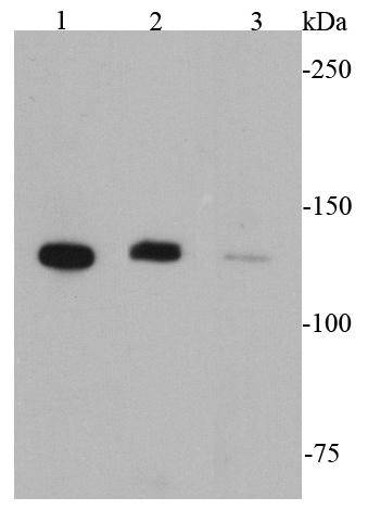
Fig1: Western blot analysis of CD31 on different cell lysates using anti-CD31 antibody at 1/1000 dilution.Application
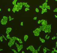
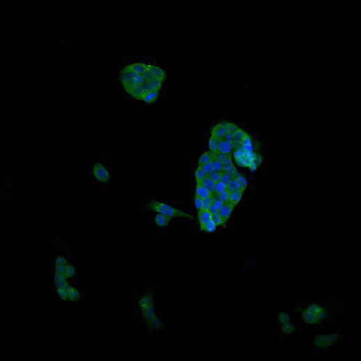
Fig2: ICC staining CD31 in Hela cells (green). Cells were fixed in paraformaldehyde, permeabilised with 0.25% Triton X100/PBS.Application
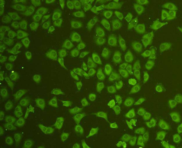
Fig3: ICC staining CD31 in HUVEC cells (green). Cells were fixed in paraformaldehyde, permeabilised with 0.25% Triton X100/PBS.Application
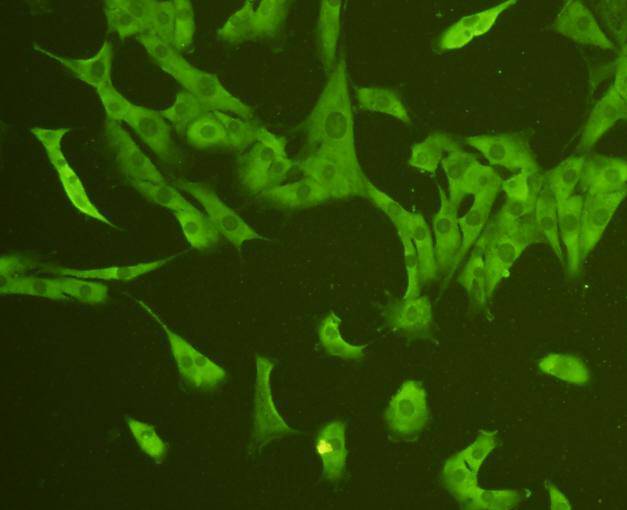
Fig4: ICC staining CD31 in NIH/3T3 cells (green). Cells were fixed in paraformaldehyde, permeabilised with 0.25% Triton X100/PBS.Application
Fig5: ICC staining CD31 in SW480 cells (green). Cells were fixed in paraformaldehyde, permeabilised with 0.25% Triton X100/PBS.Application
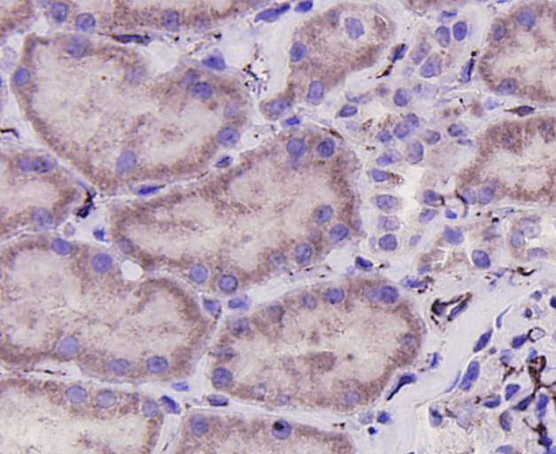
Fig6: Immunohistochemical analysis of paraffin-embedded human kidney tissue using anti-CD31 antibody. Counter stained with hematoxylin.Application
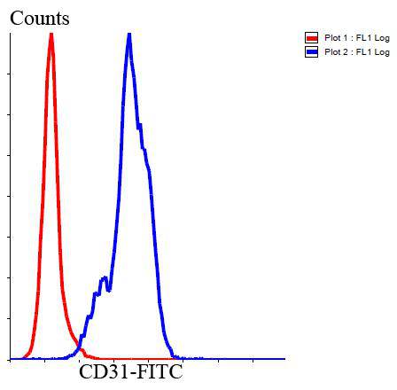
Fig7: Flow cytometric analysis of Jurkat cells with CD31 antibody at 1/100 dilution (blue) compared with an unlabelled control (cells without incubation with primary antibody; red). Goat anti rabbit IgG (FITC) was used as the secondary antibody.| Product Name | CD31 |
|---|---|
| Antibody Type | Primary Antibodies |
| Antigen Alias | Adhesion molecule antibody CD31 antibody CD31 antigen antibody CD31 EndoCAM antibody EndoCAM antibody FLJ34100 antibody FLJ58394 antibody GPIIA antibody GPIIA' antibody PECA1 antibody PECA1_HUMAN antibody Pecam 1 antibody PECAM 1 CD31 EndoCAM antibody PECAM antibody PECAM-1 antibody Pecam1 antibody Platelet and endothelial cell adhesion molecule 1 antibody Platelet endothelial cell adhesion molecule antibody Platelet/endothelial cell adhesion molecule 1 antibody |
| Product description | PECAM-1 is found on the surface of platelets, monocytes, neutrophils, and some types of T-cells, and makes up a large portion of endothelial cell intercellular junctions. The encoded protein is a member of the immunoglobulin superfamily and is likely involved in leukocyte transmigration, angiogenesis, and integrin activation. CD31 is also expressed in certain tumors, including epithelioid hemangioendothelioma, epithelioid sarcoma-like hemangioendothelioma, other vascular tumors, histiocytic malignancies, and plasmacytomas. It is rarely found in some sarcomas, such as Kaposi's sarcoma and carcinomas. In immunohistochemistry, CD31 is used primarily to demonstrate the presence of endothelial cells in histological tissue sections. This can help to evaluate the degree of tumour angiogenesis, which can imply a rapidly growing tumor |
| Immunogen | This antibody is produced by immunizing rabbits with a synthetic peptide (KLH-coupled) corresponding to C-terminal residues of human CD31. |
| Clonality | Polyclonal |
|---|---|
| Isotype | IgG |
| Host Species | Rabbit |
| Tested Applications | WBICCIHCFC |
| WB:1:1,000 ICC:1:200 IHC:1:200 FC:1:100-1:200 |
|
| Species Reactivity | HumanMouse |
| Concentration | 1mg/ml |
| Alternative Names | Adhesion molecule antibody CD31 antibody CD31 antigen antibody CD31 EndoCAM antibody EndoCAM antibody FLJ34100 antibody FLJ58394 antibody GPIIA antibody GPIIA' antibody PECA1 antibody PECA1_HUMAN antibody Pecam 1 antibody PECAM 1 CD31 EndoCAM antibody PECAM antibody PECAM-1 antibody Pecam1 antibody Platelet and endothelial cell adhesion molecule 1 antibody Platelet endothelial cell adhesion molecule antibody Platelet/endothelial cell adhesion molecule 1 antibody |
|---|---|
| Molecular Weight(MW) | 82 kDa (Observed: 130 kDa) |
| Cellular Localization | Cell membrane |
Application
Fig1: Western blot analysis of CD31 on different cell lysates using anti-CD31 antibody at 1/1000 dilution.
Application
Fig2: ICC staining CD31 in Hela cells (green). Cells were fixed in paraformaldehyde, permeabilised with 0.25% Triton X100/PBS.
Application
Fig3: ICC staining CD31 in HUVEC cells (green). Cells were fixed in paraformaldehyde, permeabilised with 0.25% Triton X100/PBS.
Application
Fig4: ICC staining CD31 in NIH/3T3 cells (green). Cells were fixed in paraformaldehyde, permeabilised with 0.25% Triton X100/PBS.
Application
Fig5: ICC staining CD31 in SW480 cells (green). Cells were fixed in paraformaldehyde, permeabilised with 0.25% Triton X100/PBS.
Application
Fig6: Immunohistochemical analysis of paraffin-embedded human kidney tissue using anti-CD31 antibody. Counter stained with hematoxylin.
Application
Fig7: Flow cytometric analysis of Jurkat cells with CD31 antibody at 1/100 dilution (blue) compared with an unlabelled control (cells without incubation with primary antibody; red). Goat anti rabbit IgG (FITC) was used as the secondary antibody.| Positive Control | THP-1,HUVEC,Hela,SW480,NIH/3T3,Jurkat,human kidney tissue |
|---|---|
| Application Notes | WB:1:1,000 ICC:1:200 IHC:1:200 FC:1:100-1:200 |
| Form | Liquid |
|---|---|
| Storage Instructions | Store at +4℃ after thawing. Aliquot store at -20℃. Avoid repeated freeze / thaw cycles. |
| Storage Buffer | 1*TBS (pH7.4), 1%BSA, 40%Glycerol. Preservative: 0.05% Sodium Azide. |


