Application
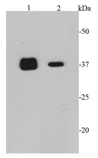
Fig1: Western blot analysis of CDK6 on different cell lysates using anti-CDK6 antibody at 1/2000 dilution. Positive control: Lane 1: K562 Lane 2: COS-1Application
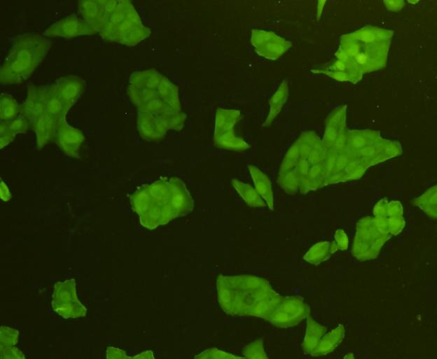
Fig2: ICC staining CDK6 in Hela cells (green). Cells were fixed in paraformaldehyde, permeabilised with 0.25% Triton X100/PBS.Application
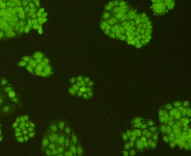
Fig3: ICC staining CDK6 in HepG2 cells (green). Cells were fixed in paraformaldehyde, permeabilised with 0.25% Triton X100/PBS.Application
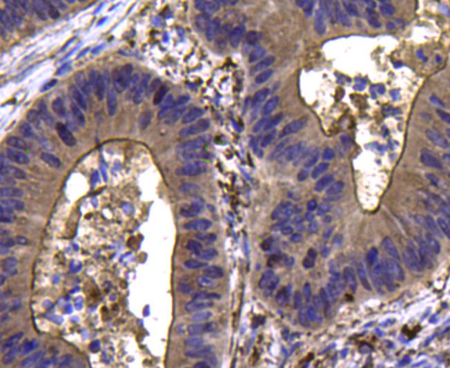
Fig4: Immunohistochemical analysis of paraffin-embedded human colon cancer tissue using anti-CDK6 antibody. Counter stained with hematoxylin.Application
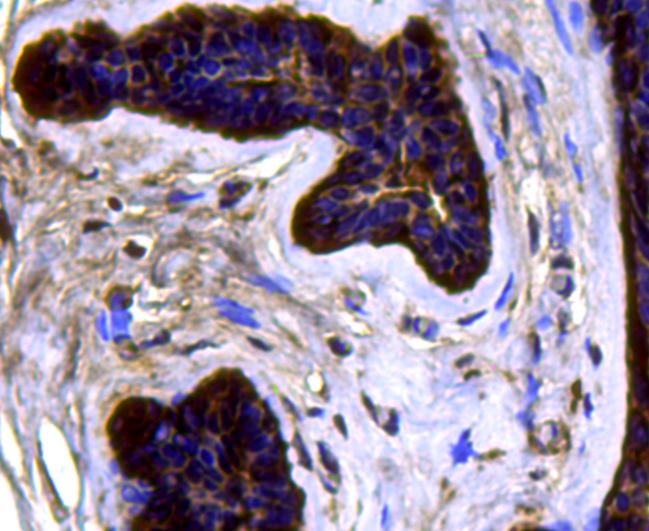
Fig5: Immunohistochemical analysis of paraffin-embedded human breast cancer tissue using anti-CDK6 antibody. Counter stained with hematoxylin.| Product Name | CDK6 |
|---|---|
| Antibody Type | Primary Antibodies |
| Antigen Alias | CDK 6 antibody CDK6 antibody CDK6_HUMAN antibody Cell division protein kinase 6 antibody Crk 2 antibody Crk2 antibody Cyclin dependent kinase 6 antibody Cyclin-dependent kinase 6 antibody MGC59692 antibody p40 antibody PLSTIRE antibody Serine/threonine protein kinase PLSTIRE antibody Serine/threonine-protein kinase PLSTIRE antibody STQTL11 antibody |
| Product description | CDK6 protein contains a catalytic core composed of a serine/threonine domain.This protein also contains an ATP-binding pocket, inhibitory and activating phosphorylation sites, a PSTAIRE-like cyclin-binding domain and an activating T-loop motif. CDK6 is involved in a positive feedback loop that activates transcription factors through a reaction cascade. CDK6 is important for the control of G1 to S phase transition. However, in recent years, new evidence proved that the presence of CDK6 is not essential for proliferation in every cell type, the cell cycle has a complex circuitry of regulation and the role of CDK6 might be more important in certain cell types than in others, where CDK4 or CDK2 can act as protein kinases compensating its role. |
| Immunogen | peptide |
| Clonality | Polyclonal |
|---|---|
| Isotype | IgG |
| Host Species | Rabbit |
| Tested Applications | WBICCIHC |
| WB:1:2,000 ICC:1:200 IHC:1:200 |
|
| Species Reactivity | HumanMouseRat |
| Concentration | 1mg/ml |
| Alternative Names | CDK 6 antibody CDK6 antibody CDK6_HUMAN antibody Cell division protein kinase 6 antibody Crk 2 antibody Crk2 antibody Cyclin dependent kinase 6 antibody Cyclin-dependent kinase 6 antibody MGC59692 antibody p40 antibody PLSTIRE antibody Serine/threonine protein kinase PLSTIRE antibody Serine/threonine-protein kinase PLSTIRE antibody STQTL11 antibody |
|---|---|
| Molecular Weight(MW) | 37 kDa |
| Cellular Localization | Cytoplasm, nucleus. |
| SwissProt ID | Q00534 |
|---|
Application
Fig1: Western blot analysis of CDK6 on different cell lysates using anti-CDK6 antibody at 1/2000 dilution. Positive control: Lane 1: K562 Lane 2: COS-1
Application
Fig2: ICC staining CDK6 in Hela cells (green). Cells were fixed in paraformaldehyde, permeabilised with 0.25% Triton X100/PBS.
Application
Fig3: ICC staining CDK6 in HepG2 cells (green). Cells were fixed in paraformaldehyde, permeabilised with 0.25% Triton X100/PBS.
Application
Fig4: Immunohistochemical analysis of paraffin-embedded human colon cancer tissue using anti-CDK6 antibody. Counter stained with hematoxylin.
Application
Fig5: Immunohistochemical analysis of paraffin-embedded human breast cancer tissue using anti-CDK6 antibody. Counter stained with hematoxylin.| Positive Control | Hela, HepG2, COS-1, K562, human colon cancer tissue, human breast cancer tissue. |
|---|---|
| Application Notes | WB:1:2,000 ICC:1:200 IHC:1:200 |
| Form | Liquid |
|---|---|
| Storage Instructions | Store at +4℃ after thawing. Aliquot store at -20℃ or -80℃. Avoid repeated freeze / thaw cycles. |
| Storage Buffer | 1*TBS (pH7.4), 1%BSA, 40%Glycerol. Preservative: 0.05% Sodium Azide. |


