Application
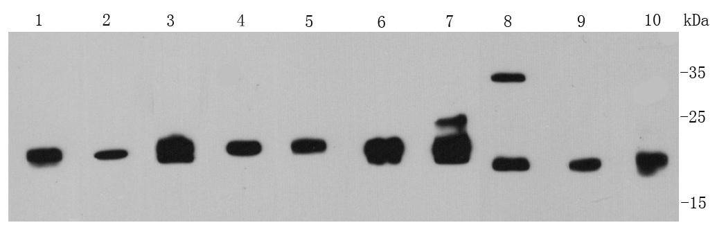
Fig1: Western blot analysis of MGMT on different cell lysates using anti- MGMT antibody at 1/1000 dilution. Positive control: Lane 1: Hela Lane 2: MCF-7 Lane 3: Daudi Lane 4: Raji Lane 5: SK-BR-3 Lane 6: SKOV3 Lane 7: Human liver Lane 8: Human kidney Lane 9: Human thymus Lane 10: Human placentaeApplication
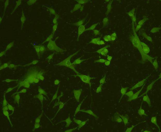
Fig2: ICC staining MGMT in A172 cells (green). Cells were fixed in paraformaldehyde, permeabilised with 0.25% Triton X100/PBS.Application
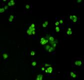
Fig3: ICC staining MGMT in SK-BR-3 cells (green). Cells were fixed in paraformaldehyde, permeabilised with 0.25% Triton X100/PBS.Application
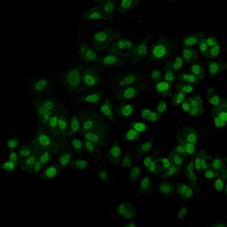
Fig4: ICC staining MGMT in MCF-7 cells (green). Cells were fixed in paraformaldehyde, permeabilised with 0.25% Triton X100/PBS.Application
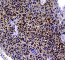
Fig5: Immunohistochemical analysis of paraffin-embedded human tonsil tissue using anti-MGMT antibody. Counter stained with hematoxylin.Application
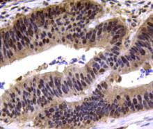
Fig6: Immunohistochemical analysis of paraffin-embedded human colon carcinoma tissue using anti-MGMT antibody. Counter stained with hematoxylin.| Product Name | MGMT |
|---|---|
| Antibody Type | Primary Antibodies |
| Antigen Alias | 6 O methylguanine DNA methyltransferase antibody 6-O-methylguanine-DNA methyltransferase antibody Agat antibody AGT antibody AI267024 antibody EC 2.1.1.63 antibody Methylated DNA protein cysteine methyltransferase antibody Methylated-DNAprotein-cysteine methyltransferase antibody Methylguanine DNA methyltransferase antibody MGC107020 antibody MGMT antibody MGMT_HUMAN antibody O 6 methylguanine DNA alkyltransferase antibody O 6 methylguanine DNA methyltransferase antibody O-6-methylguanine-DNA methyltransferase antibody O-6-methylguanine-DNA-alkyltransferase antibody |
| Product description | MGMT (O-6-methylguanine-DNA methyltransferase) is a DNA repair enzyme that participates in a suicide reaction that specifically removes methyl or alkyl groups from the O (6) position of guanine, restoring guanine to its normal form without causing DNA breaks. MGMT protects cells from alkylating toxins, and is an important factor in drug resistance to alkylating therapeutic agents. It is ubiquitously expressed in normal human tissues and is overexpressed in many types of human tumors, but epigenetically silenced in other tumors. MGMT silencing is a marker associated with poor prognosis, but is a good predictive marker for response to alkylating agent chemotherapy. |
| Immunogen | peptide |
| Clonality | Polyclonal |
|---|---|
| Isotype | IgG |
| Host Species | Rabbit |
| Tested Applications | WBICCIHCFC |
| WB:1:500-1:1,000 ICC:1:200 IHC:1:200 FC:1:100: |
|
| Species Reactivity | Human |
| Concentration | 1mg/ml |
| Alternative Names | 6 O methylguanine DNA methyltransferase antibody 6-O-methylguanine-DNA methyltransferase antibody Agat antibody AGT antibody AI267024 antibody EC 2.1.1.63 antibody Methylated DNA protein cysteine methyltransferase antibody Methylated-DNAprotein-cysteine methyltransferase antibody Methylguanine DNA methyltransferase antibody MGC107020 antibody MGMT antibody MGMT_HUMAN antibody O 6 methylguanine DNA alkyltransferase antibody O 6 methylguanine DNA methyltransferase antibody O-6-methylguanine-DNA methyltransferase antibody O-6-methylguanine-DNA-alkyltransferase antibody |
|---|---|
| Molecular Weight(MW) | 22 kDa |
| Cellular Localization | Nucleus |
| SwissProt ID | P16455 |
|---|
Application
Fig1: Western blot analysis of MGMT on different cell lysates using anti- MGMT antibody at 1/1000 dilution. Positive control: Lane 1: Hela Lane 2: MCF-7 Lane 3: Daudi Lane 4: Raji Lane 5: SK-BR-3 Lane 6: SKOV3 Lane 7: Human liver Lane 8: Human kidney Lane 9: Human thymus Lane 10: Human placentae
Application
Fig2: ICC staining MGMT in A172 cells (green). Cells were fixed in paraformaldehyde, permeabilised with 0.25% Triton X100/PBS.
Application
Fig3: ICC staining MGMT in SK-BR-3 cells (green). Cells were fixed in paraformaldehyde, permeabilised with 0.25% Triton X100/PBS.
Application
Fig4: ICC staining MGMT in MCF-7 cells (green). Cells were fixed in paraformaldehyde, permeabilised with 0.25% Triton X100/PBS.
Application
Fig5: Immunohistochemical analysis of paraffin-embedded human tonsil tissue using anti-MGMT antibody. Counter stained with hematoxylin.
Application
Fig6: Immunohistochemical analysis of paraffin-embedded human colon carcinoma tissue using anti-MGMT antibody. Counter stained with hematoxylin.| Positive Control | MCF-7, Hela, Dauai, Raji, A172,human kidney tissue, human liver tissue, human thymus tissue, human placentae tissue, Jurkat,SK-BR-3 ,SKOV3,human tonsil tissuehuman colon carcinoma tissue. |
|---|---|
| Application Notes | WB:1:500-1:1,000 ICC:1:200 IHC:1:200 FC:1:100: |
| Form | Liquid |
|---|---|
| Storage Instructions | Store at +4℃ after thawing. Aliquot store at -20℃ or -80℃. Avoid repeated freeze / thaw cycles. |
| Storage Buffer | 1*TBS (pH7.4), 1%BSA, 40%Glycerol. Preservative: 0.05% Sodium Azide. |


