Application
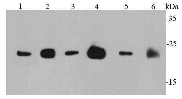
Fig1: Western blot analysis of Ras on different cell lysates using anti-Ras antibody at 1/1000 dilution. Positive control: Lane 1: PC12 Lane 2: MCF-7 Lane 3: 293T Lane 4: Mouse brain Lane 5: Mouse intestine Lane 6: Rat brainApplication
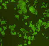
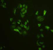
Fig2: ICC staining Ras in HCT116 cells (green). Cells were fixed in paraformaldehyde, permeabilised with 0.25% Triton X100/PBS.Application
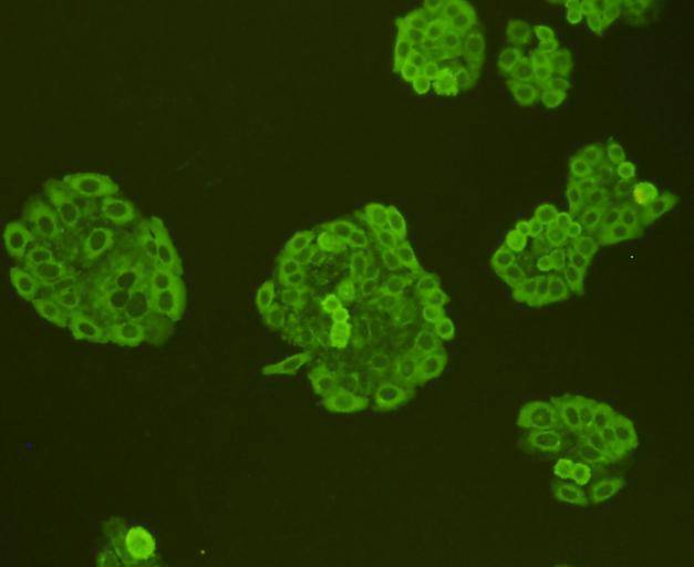
Fig3: ICC staining Ras in HepG2 cells (green). Cells were fixed in paraformaldehyde, permeabilised with 0.25% Triton X100/PBS.Application
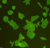
Fig4: ICC staining Ras in Hela cells (green). Cells were fixed in paraformaldehyde, permeabilised with 0.25% Triton X100/PBS.Application
Fig5: ICC staining Ras in MCF-7 cells (green). Cells were fixed in paraformaldehyde, permeabilised with 0.25% Triton X100/PBS.Application
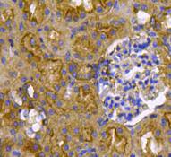
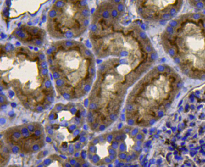
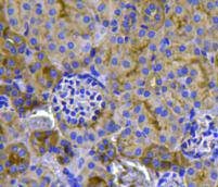
Fig6: Immunohistochemical analysis of paraffin-embedded mouse kidney tissue using anti-Ras antibody. Counter stained with hematoxylin.Application
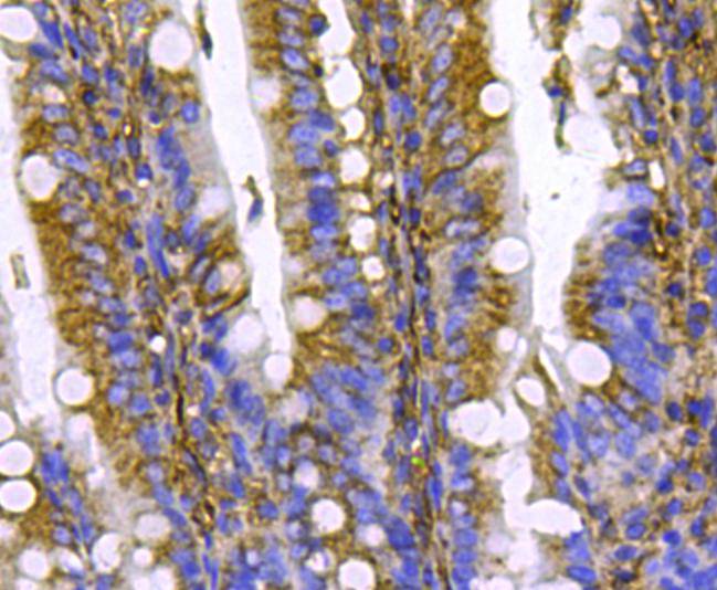
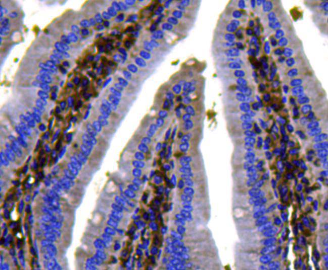
Fig7: Immunohistochemical analysis of paraffin-embedded rat small intestine tissue using anti-Ras antibody. Counter stained with hematoxylin.Application
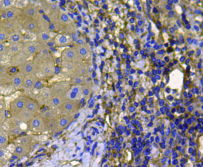
Fig8: Immunohistochemical analysis of paraffin-embedded human liver cancer tissue using anti-Ras antibody. Counter stained with hematoxylin.Application
Fig9: Immunohistochemical analysis of paraffin-embedded human kidney muscle tissue using anti-Ras antibody. Counter stained with hematoxylin.Application
Fig10: Immunohistochemical analysis of paraffin-embedded mouse kidney tissue using anti-Ras antibody. Counter stained with hematoxylin.Application
Fig11: Immunohistochemical analysis of paraffin-embedded mouse small intestine tissue using anti-Ras antibody. Counter stained with hematoxylin.| Product Name | Ras |
|---|---|
| Antibody Type | Primary Antibodies |
| Antigen Alias | C-BAS/HAS antibody c-H-ras antibody C-HA-RAS1 antibody CTLO antibody GTPase HRas antibody GTPase KRas antibody GTPase NRas antibody H-Ras-1 antibody H-RASIDX antibody Ha-Ras antibody HAMSV antibody HRAS antibody HRAS1 antibody K RAS2A antibody K RAS2B antibody K RAS4A antibody K RAS4B antibody K-RAS antibody KRAS antibody KRAS1 antibody KRAS2 antibody N-RAS antibody N-terminally processed antibody NRAS antibody NRAS1 antibody p21ras antibody RASH_HUMAN antibody RASH1 antibody RASK2 antibody Transforming protein p21 antibody v Ha ras Harvey rat sarcoma viral oncogene homolog antibody v Ki ras2 Kirsten rat sarcoma viral oncogene homolog antibody v ras neuroblastoma RAS viral oncogene homolog antibody |
| Product description | Ras superfamily is a protein superfamily of small GTPases, which are all related, to a degree, to the Ras protein subfamily (the key human members of which are KRAS, NRAS, and HRAS). Receptor tyrosine kinases and G protein-coupled receptors activate Ras, which then stimulates the Raf-MEK-MAPK pathway. GTPase-activating proteins (GAP) normally facilitate the inactivation of Ras. However, research studies have shown that in 30% of human tumors, point mutations in Ras prevent the GAP-mediated inhibition of this pathway. The most common oncogenic Ras mutation found in tumors is Gly12 to Asp12 (G12D), which prevents Ras inactivation, possibly by increasing the overall rigidity of the protein. This antibody is predicted to react with H-Ras, N-Ras and K-Ras. |
| Immunogen | peptide |
| Clonality | Polyclonal |
|---|---|
| Isotype | IgG |
| Host Species | Rabbit |
| Tested Applications | WBICCIHCFC |
| WB:1:1,000 ICC:1:200 IHC:1:200 FC:1:100-1:200 |
|
| Species Reactivity | HumanMouseRat |
| Concentration | 1mg/ml |
| Alternative Names | C-BAS/HAS antibody c-H-ras antibody C-HA-RAS1 antibody CTLO antibody GTPase HRas antibody GTPase KRas antibody GTPase NRas antibody H-Ras-1 antibody H-RASIDX antibody Ha-Ras antibody HAMSV antibody HRAS antibody HRAS1 antibody K RAS2A antibody K RAS2B antibody K RAS4A antibody K RAS4B antibody K-RAS antibody KRAS antibody KRAS1 antibody KRAS2 antibody N-RAS antibody N-terminally processed antibody NRAS antibody NRAS1 antibody p21ras antibody RASH_HUMAN antibody RASH1 antibody RASK2 antibody Transforming protein p21 antibody v Ha ras Harvey rat sarcoma viral oncogene homolog antibody v Ki ras2 Kirsten rat sarcoma viral oncogene homolog antibody v ras neuroblastoma RAS viral oncogene homolog antibody |
|---|---|
| Molecular Weight(MW) | 21 kDa |
| Cellular Localization | Cytoplasmic side, cell membrane, lipid-anchor, golgi apparatus membrane |
Application
Fig1: Western blot analysis of Ras on different cell lysates using anti-Ras antibody at 1/1000 dilution. Positive control: Lane 1: PC12 Lane 2: MCF-7 Lane 3: 293T Lane 4: Mouse brain Lane 5: Mouse intestine Lane 6: Rat brain
Application
Fig2: ICC staining Ras in HCT116 cells (green). Cells were fixed in paraformaldehyde, permeabilised with 0.25% Triton X100/PBS.
Application
Fig3: ICC staining Ras in HepG2 cells (green). Cells were fixed in paraformaldehyde, permeabilised with 0.25% Triton X100/PBS.
Application
Fig4: ICC staining Ras in Hela cells (green). Cells were fixed in paraformaldehyde, permeabilised with 0.25% Triton X100/PBS.
Application
Fig5: ICC staining Ras in MCF-7 cells (green). Cells were fixed in paraformaldehyde, permeabilised with 0.25% Triton X100/PBS.
Application
Fig6: Immunohistochemical analysis of paraffin-embedded mouse kidney tissue using anti-Ras antibody. Counter stained with hematoxylin.
Application
Fig7: Immunohistochemical analysis of paraffin-embedded rat small intestine tissue using anti-Ras antibody. Counter stained with hematoxylin.
Application
Fig8: Immunohistochemical analysis of paraffin-embedded human liver cancer tissue using anti-Ras antibody. Counter stained with hematoxylin.
Application
Fig9: Immunohistochemical analysis of paraffin-embedded human kidney muscle tissue using anti-Ras antibody. Counter stained with hematoxylin.
Application
Fig10: Immunohistochemical analysis of paraffin-embedded mouse kidney tissue using anti-Ras antibody. Counter stained with hematoxylin.
Application
Fig11: Immunohistochemical analysis of paraffin-embedded mouse small intestine tissue using anti-Ras antibody. Counter stained with hematoxylin.| Positive Control | PC12, MCF-7, 293T, Hela, HepG2, mouse brain tissue, mouse intestines tissue, rat brain tissue, human kidney tissue, mouse kidney tissue, mouse small intestine tissue, mouse pancreas tissue, rat kidney tissue, rat small intestine tissue, human liver cancer tissue. |
|---|---|
| Application Notes | WB:1:1,000 ICC:1:200 IHC:1:200 FC:1:100-1:200 |
| Form | Liquid |
|---|---|
| Storage Instructions | Store at +4℃ after thawing. Aliquot store at -20℃ or -80℃. Avoid repeated freeze / thaw cycles. |
| Storage Buffer | 1*TBS (pH7.4), 1%BSA, 40%Glycerol. Preservative: 0.05% Sodium Azide. |


