Application
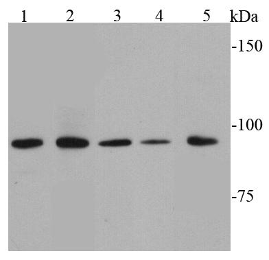
Fig1: Western blot analysis of STAT6 on different cell lysates using anti-STAT6 antibody at 1/1000 dilution. Positive control: Lane 1: Raji Lane 2: Daudi Lane 3: Hela Lane 4: Ags Lane 5: JurkatApplication
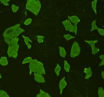
Fig2: ICC staining STAT6 in Hela cells (green). Cells were fixed in paraformaldehyde, permeabilised with 0.25% Triton X100/PBS.Application
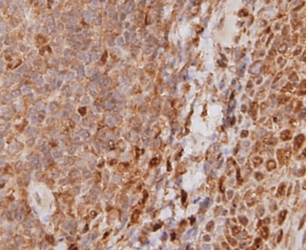
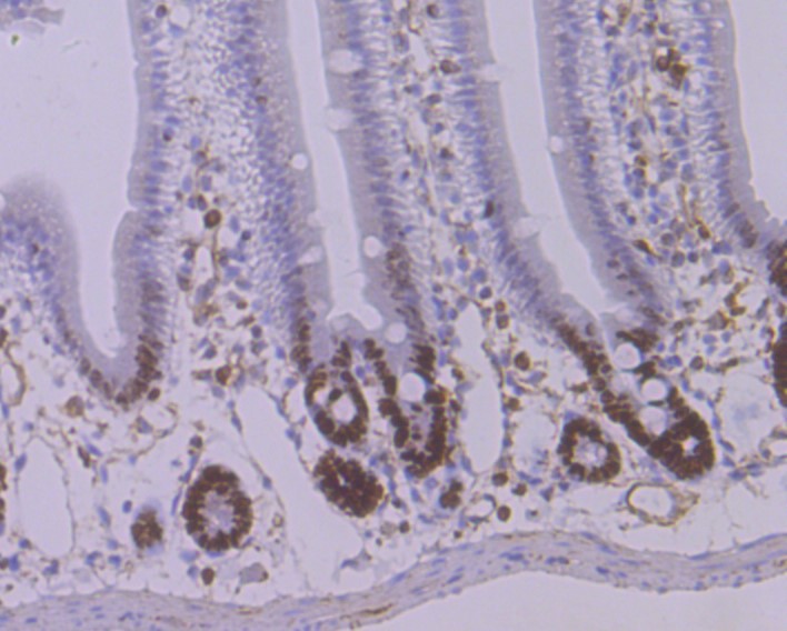
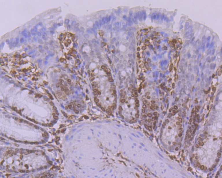
Fig3: Immunohistochemical analysis of paraffin-embedded human tonsil tissue using anti-STAT6 antibody. Counter stained with hematoxylin.Application
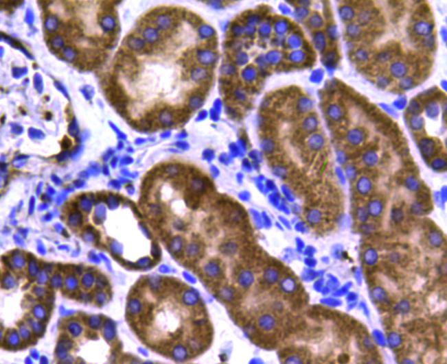
Fig4: Immunohistochemical analysis of paraffin-embedded human kidney tissue using anti-STAT6 antibody. Counter stained with hematoxylin.Application
Fig5: Immunohistochemical analysis of paraffin-embedded human gastric carcinoma tissue using anti-STAT6 antibody. Counter stained with hematoxylin.Application
Fig6: Flow cytometric analysis of Hela cells with ATAT6 antibody at 1/100 dilution (blue) compared with an unlabelled control (cells without incubation with primary antibody; red). Goat anti rabbit IgG (FITC) was used as the secondary antibody.| Product Name | STAT6 |
|---|---|
| Antibody Type | Primary Antibodies |
| Antigen Alias | 12S1644 antibody D12S1644 antibody IL 4 STAT antibody IL-4 Stat antibody IL4 STAT antibody Interleukin 4 Induced antibody Interleukin 4 Induced Transcription Factor IL4 STAT antibody Signal transducer and activator of transcription 6 antibody Signal Transducer And Activator Of Transcription 6 Interleukin 4 Induced antibody Signal Transducer And Activator Of Transcription 6 Nirs Variant 1 antibody Signal transducer and activator of transcription 6, interleukin 4 induced antibody STAT 6 antibody STAT interleukin4 induced antibody STAT, interleukin4 induced antibody Stat6 antibody STAT6_HUMAN antibody STAT6B antibody STAT6C antibody Transcription factor IL 4 STAT antibody |
| Product description | In response to cytokines and growth factors, STAT family members are phosphorylated by the receptor associated kinases, and then form homo- or heterodimers that translocate to the cell nucleus where they act as transcription activators. This protein STAT6 plays a central role in exerting IL4 mediated biological responses. It is found to induce the expression of BCL2L1/BCL-X(L), which is responsible for the anti-apoptotic activity of IL4. Knockout studies in mice suggested the roles of this gene in differentiation of T helper 2 (Th2), expression of cell surface markers, and class switch of immunoglobulins. |
| Immunogen | peptide |
| Clonality | Polyclonal |
|---|---|
| Isotype | IgG |
| Host Species | Rabbit |
| Tested Applications | WBICCIHCFC |
| WB:1:1,000 ICC:1:200 IHC:1:200 FC:1:100-1:200 |
|
| Species Reactivity | Human |
| Concentration | 1mg/ml |
| Alternative Names | 12S1644 antibody
D12S1644 antibody
IL 4 STAT antibody
IL-4 Stat antibody
IL4 STAT antibody
Interleukin 4 Induced antibody
Interleukin 4 Induced Transcription Factor IL4 STAT antibody
Signal transducer and activator of transcription 6 antibody
Signal Transducer And Activator Of Transcription 6 Interleukin 4 Induced antibody
Signal Transducer And Activator Of Transcription 6 Nirs Variant 1 antibody
Signal transducer and activator of transcription 6 interleukin 4 induced antibody STAT 6 antibody STAT interleukin4 induced antibody STAT interleukin4 induced antibody Stat6 antibody STAT6_HUMAN antibody STAT6B antibody STAT6C antibody Transcription factor IL 4 STAT antibody |
|---|---|
| Molecular Weight(MW) | 94 kDa |
| Cellular Localization | Cytoplasm, nucleus. |
| SwissProt ID | P42226 |
|---|
Application
Fig1: Western blot analysis of STAT6 on different cell lysates using anti-STAT6 antibody at 1/1000 dilution. Positive control: Lane 1: Raji Lane 2: Daudi Lane 3: Hela Lane 4: Ags Lane 5: Jurkat
Application
Fig2: ICC staining STAT6 in Hela cells (green). Cells were fixed in paraformaldehyde, permeabilised with 0.25% Triton X100/PBS.
Application
Fig3: Immunohistochemical analysis of paraffin-embedded human tonsil tissue using anti-STAT6 antibody. Counter stained with hematoxylin.
Application
Fig4: Immunohistochemical analysis of paraffin-embedded human kidney tissue using anti-STAT6 antibody. Counter stained with hematoxylin.
Application
Fig5: Immunohistochemical analysis of paraffin-embedded human gastric carcinoma tissue using anti-STAT6 antibody. Counter stained with hematoxylin.
Application
Fig6: Flow cytometric analysis of Hela cells with ATAT6 antibody at 1/100 dilution (blue) compared with an unlabelled control (cells without incubation with primary antibody; red). Goat anti rabbit IgG (FITC) was used as the secondary antibody.| Positive Control | Jurkat, Raji, Daudi, Hela, Ags, human tosnsil tissue, human kidney tissue, human gastric carcinoma tissue. |
|---|---|
| Application Notes | WB:1:1,000 ICC:1:200 IHC:1:200 FC:1:100-1:200 |
| Form | Liquid |
|---|---|
| Storage Instructions | Store at +4℃ after thawing. Aliquot store at -20℃ or -80℃. Avoid repeated freeze / thaw cycles. |
| Storage Buffer | 1*TBS (pH7.4), 1%BSA, 40%Glycerol. Preservative: 0.05% Sodium Azide. |


