Application
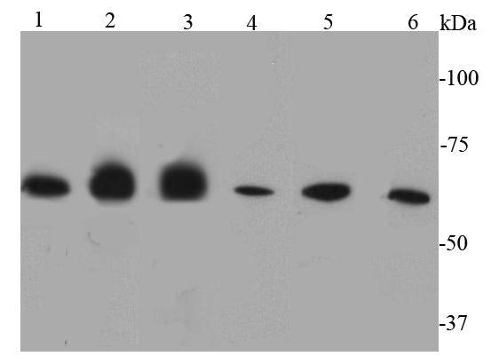
Fig1: Western blot analysis of Caveolin-1 on different lysates using anti-Caveolin-1 antibody at 1/500 dilution.Positive control:Lane 1: F9 Lane 2: A549Lane 3: Mouse lung Lane 4: Mouse liverLane 5: Human lung Lane 6: HelaLane 7: HepG2 Lane 8: JurkatApplication
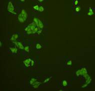
Fig2: ICC staining catalase in Hela cells (green). Cells were fixed in paraformaldehyde, permeabilised with 0.25% Triton X100/PBS.Application
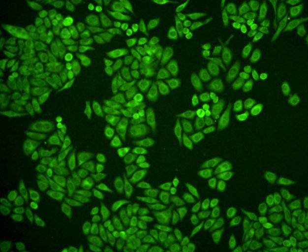
Fig3: ICC staining catalase in HepG2 cells (green). Cells were fixed in paraformaldehyde, permeabilised with 0.25% Triton X100/PBS.Application
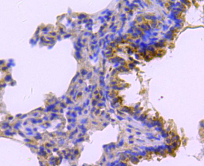
Fig4: Immunohistochemical analysis of paraffin-embedded rat lung tissue using anti-catalase antibody. Counter stained with hematoxylin.Application
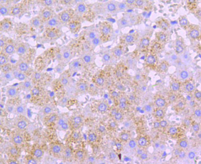
Fig5: Immunohistochemical analysis of paraffin-embedded rat liver tissue using anti-catalase antibody. Counter stained with hematoxylin.Application
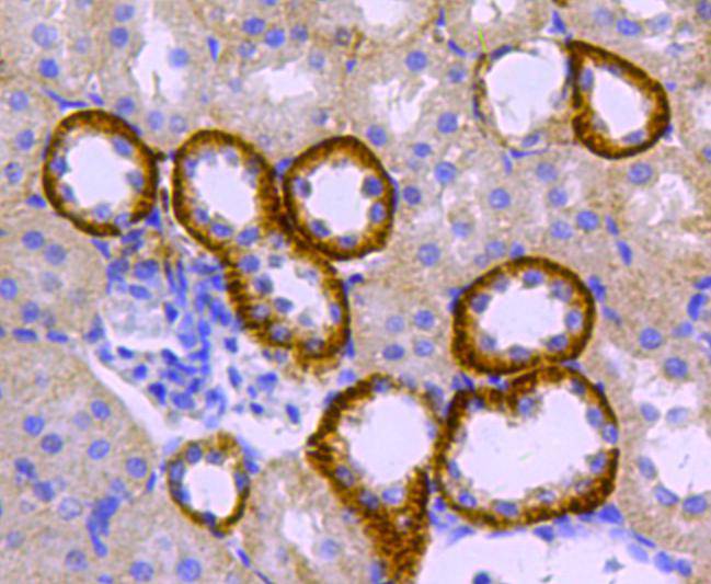
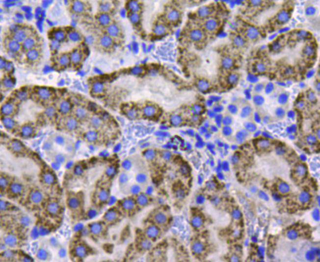
Fig6: Immunohistochemical analysis of paraffin-embedded rat kidney tissue using anti-catalase antibody. Counter stained with hematoxylin.Application
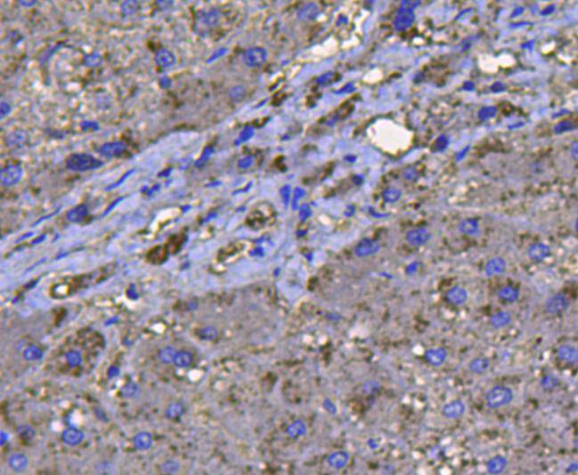
Fig7: Immunohistochemical analysis of paraffin-embedded human liver tissue using anti-catalase antibody. Counter stained with hematoxylin.Application
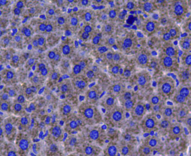
Fig8: Immunohistochemical analysis of paraffin-embedded mouse liver tissue using anti-catalase antibody. Counter stained with hematoxylin.Application
Fig9: Immunohistochemical analysis of paraffin-embedded mouse kidney tissue using anti-catalase antibody. Counter stained with hematoxylin.| Product Name | Catalase |
|---|---|
| Antibody Type | Primary Antibodies |
| Antigen Alias | Cas1 antibody CAT antibody CATA_HUMAN antibody Catalase antibody Cs1 antibody MGC138422 antibody MGC138424 antibody |
| Product description | Hydrogen peroxide is a harmful byproduct of many normal metabolic processes; to prevent damage to cells and tissues, it must be quickly converted into other, less dangerous substances. To this end, catalase is frequently used by cells to rapidly catalyze the decomposition of hydrogen peroxide into less-reactive gaseous oxygen and water molecules. Catalase is usually located in a cellular, bipolar environment organelle called the peroxisome. All known animals use catalase in every organ, with particularly high concentrations occurring in the liver. Catalase promotes growth of cells including T-cells, B-cells, myeloid leukemia cells, melanoma cells, mastocytoma cells and normal and transformed fibroblast cells. |
| Immunogen | Synthetic peptide (KLH-coupled) within human Caveolin-1 110-180aa. |
| Clonality | Polyclonal |
|---|---|
| Isotype | IgG |
| Host Species | Rabbit |
| Tested Applications | WBICCIHC |
| WB:1:1,000 ICC:1:200 IHC:1:200 |
|
| Species Reactivity | HumanMouseRat |
| Concentration | 1mg/ml |
| Alternative Names | Cas1 antibody CAT antibody CATA_HUMAN antibody Catalase antibody Cs1 antibody MGC138422 antibody MGC138424 antibody |
|---|---|
| Molecular Weight(MW) | 60 kDa |
| Cellular Localization | Peroxisome |
Application
Fig1: Western blot analysis of Caveolin-1 on different lysates using anti-Caveolin-1 antibody at 1/500 dilution.Positive control:Lane 1: F9 Lane 2: A549Lane 3: Mouse lung Lane 4: Mouse liverLane 5: Human lung Lane 6: HelaLane 7: HepG2 Lane 8: Jurkat
Application
Fig2: ICC staining catalase in Hela cells (green). Cells were fixed in paraformaldehyde, permeabilised with 0.25% Triton X100/PBS.
Application
Fig3: ICC staining catalase in HepG2 cells (green). Cells were fixed in paraformaldehyde, permeabilised with 0.25% Triton X100/PBS.
Application
Fig4: Immunohistochemical analysis of paraffin-embedded rat lung tissue using anti-catalase antibody. Counter stained with hematoxylin.
Application
Fig5: Immunohistochemical analysis of paraffin-embedded rat liver tissue using anti-catalase antibody. Counter stained with hematoxylin.
Application
Fig6: Immunohistochemical analysis of paraffin-embedded rat kidney tissue using anti-catalase antibody. Counter stained with hematoxylin.
Application
Fig7: Immunohistochemical analysis of paraffin-embedded human liver tissue using anti-catalase antibody. Counter stained with hematoxylin.
Application
Fig8: Immunohistochemical analysis of paraffin-embedded mouse liver tissue using anti-catalase antibody. Counter stained with hematoxylin.
Application
Fig9: Immunohistochemical analysis of paraffin-embedded mouse kidney tissue using anti-catalase antibody. Counter stained with hematoxylin.| Positive Control | Hela, HepG2, A431, mouse liver tissue, mouse kidney tissue, human liver tissue, rat kidney tissue, rat liver tissue, rat lung tissue |
|---|---|
| Application Notes | WB:1:1,000 ICC:1:200 IHC:1:200 |
| Form | Liquid |
|---|---|
| Storage Instructions | Store at +4℃ after thawing. Aliquot store at -20℃. Avoid repeated freeze / thaw cycles. |
| Storage Buffer | 1*TBS (pH7.4), 1%BSA, 40%Glycerol. Preservative: 0.05% Sodium Azide. |


