Application
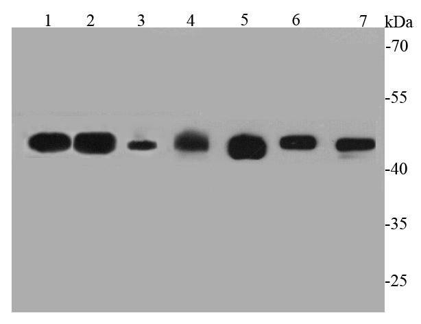
Fig1: Western blot analysis of GAP43 on different cell lysates using anti-GAP43 antibody at 1/1000 dilution.Positive control:Lane 1: Rat brainLane 2: Mouse brainLane 3: Mouse heartLane 4: Human skeletal muscleLane 5: N2ALane 6: A172Lane 7: Human heartApplication
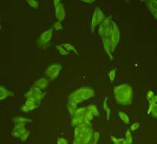
Fig2: ICC staining GAP43 in Hela cells (green). Cells were fixed in paraformaldehyde, permeabilised with 0.25% Triton X100/PBS.Application
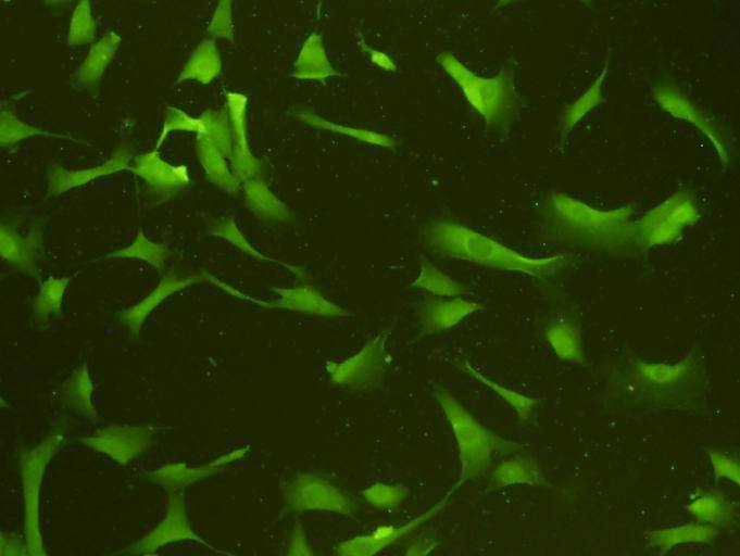
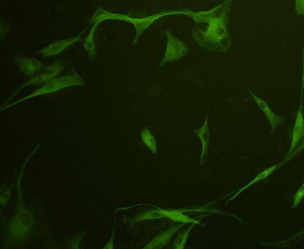
Fig3: ICC staining GAP43 in NIH/3T3 cells (green). Cells were fixed in paraformaldehyde, permeabilised with 0.25% Triton X100/PBS.Application
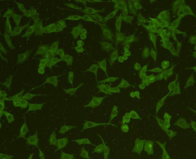
Fig4: ICC staining GAP43 in SHG-44 cells (green). Cells were fixed in paraformaldehyde, permeabilised with 0.25% Triton X100/PBS.Application
Fig5: ICC staining GAP43 in A172 cells (green). Cells were fixed in paraformaldehyde, permeabilised with 0.25% Triton X100/PBS.Application
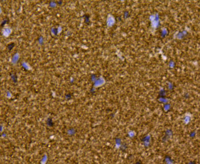
Fig6: Immunohistochemical analysis of paraffin-embedded rat brain tissue using anti-GAP43 antibody. Counter stained with hematoxylin.Application
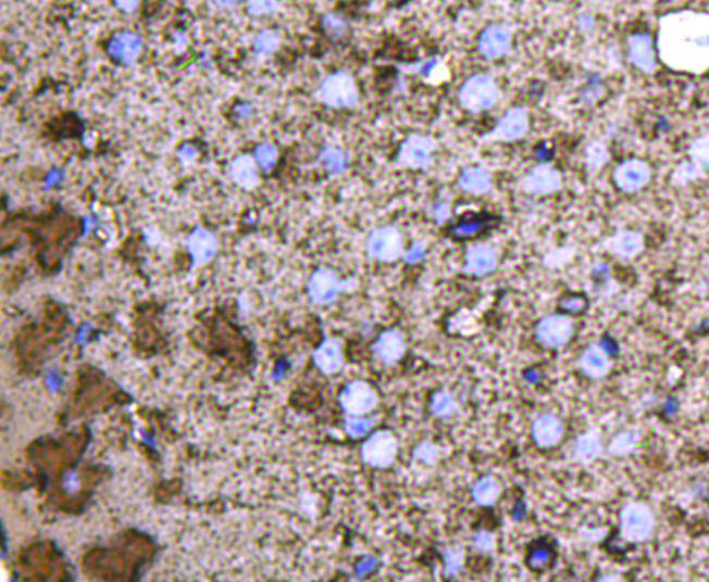
Fig7: Immunohistochemical analysis of paraffin-embedded mouse brain tissue using anti-GAP43 antibody. Counter stained with hematoxylin.| Product Name | GAP43 |
|---|---|
| Antibody Type | Primary Antibodies |
| Antigen Alias | Axonal membrane protein GAP 43 antibody Axonal membrane protein GAP-43 antibody B 50 antibody Calmodulin binding protein P 57 antibody F1 antibody GAP 43 antibody GAP43 antibody Growth Associated Protein 43 antibody Growth-associated protein 43 antibody Nerve Growth Related Peptide antibody Nerve growth related peptide GAP43 antibody NEUM_HUMAN antibody Neural phosphoprotein B 50 antibody Neural phosphoprotein B-50 antibody Neuromodulin antibody Neuron growth associated protein 43 antibody PP46 antibody Protein F1 antibody QtrA-11580 antibody QtrA-13071 antibody |
| Product description | GAP43, is a nervous tissue-specific cytoplasmic protein that can be attached to the membrane via a dual palmitoylation sequence on cysteines 3 and 4. This sequence targets GAP43 to lipid rafts. It is a major protein kinase C (PKC) substrate and is considered to play a key role in neurite formation, regeneration, and plasticity. The role of GAP-43 in CNS development is not limited to effects on axons: It is also a component of the centrosome, and differentiating neurons that do not express GAP-43 show mislocalization of the centrosome and mitotic spindles, particularly in neurogenic cell divisions. As a consequence, in the cerebellum, the neuronal precursor pool fails to expand normally and the cerebellum is significantly smaller. |
| Immunogen | peptide |
| Clonality | Polyclonal |
|---|---|
| Isotype | IgG |
| Host Species | Rabbit |
| Tested Applications | WBICCIHCFC |
| WB:1:1,000 ICC:1:200 IHC:1:200 FC:1:100-1:200 |
|
| Species Reactivity | HumanMouseRat |
| Concentration | 1mg/ml |
| Alternative Names | Axonal membrane protein GAP 43 antibody Axonal membrane protein GAP-43 antibody B 50 antibody Calmodulin binding protein P 57 antibody F1 antibody GAP 43 antibody GAP43 antibody Growth Associated Protein 43 antibody Growth-associated protein 43 antibody Nerve Growth Related Peptide antibody Nerve growth related peptide GAP43 antibody NEUM_HUMAN antibody Neural phosphoprotein B 50 antibody Neural phosphoprotein B-50 antibody Neuromodulin antibody Neuron growth associated protein 43 antibody PP46 antibody Protein F1 antibody QtrA-11580 antibody QtrA-13071 antibody |
|---|---|
| Molecular Weight(MW) | 43 kDa |
| Cellular Localization | Cell membrane. |
| SwissProt ID | P06837 |
|---|
Application
Fig1: Western blot analysis of GAP43 on different cell lysates using anti-GAP43 antibody at 1/1000 dilution.Positive control:Lane 1: Rat brainLane 2: Mouse brainLane 3: Mouse heartLane 4: Human skeletal muscleLane 5: N2ALane 6: A172Lane 7: Human heart
Application
Fig2: ICC staining GAP43 in Hela cells (green). Cells were fixed in paraformaldehyde, permeabilised with 0.25% Triton X100/PBS.
Application
Fig3: ICC staining GAP43 in NIH/3T3 cells (green). Cells were fixed in paraformaldehyde, permeabilised with 0.25% Triton X100/PBS.
Application
Fig4: ICC staining GAP43 in SHG-44 cells (green). Cells were fixed in paraformaldehyde, permeabilised with 0.25% Triton X100/PBS.
Application
Fig5: ICC staining GAP43 in A172 cells (green). Cells were fixed in paraformaldehyde, permeabilised with 0.25% Triton X100/PBS.
Application
Fig6: Immunohistochemical analysis of paraffin-embedded rat brain tissue using anti-GAP43 antibody. Counter stained with hematoxylin.
Application
Fig7: Immunohistochemical analysis of paraffin-embedded mouse brain tissue using anti-GAP43 antibody. Counter stained with hematoxylin.| Positive Control | Hela, NIH/3T3, N2A, A172, SHG-44, mouse brain tissue, rat brain tissue, human skeletal muscle tissue, human heart tissue, mouse heart tissue. |
|---|---|
| Application Notes | WB:1:1,000 ICC:1:200 IHC:1:200 FC:1:100-1:200 |
| Form | Liquid |
|---|---|
| Storage Instructions | Store at +4℃ after thawing. Aliquot store at -20℃ or -80℃. Avoid repeated freeze / thaw cycles. |
| Storage Buffer | 1*TBS (pH7.4), 1%BSA, 40%Glycerol. Preservative: 0.05% Sodium Azide. |


