
Application
Fig1: Western blot analysis of PDI on different cell lysates using anti- PDI antibody at 1/1000 dilution. Positive control: Lane 1: Hela Lane 2: MCF-7 Lane 3: PANC Lane 4: HepG2 Lane 5: HUVEC Lane 6: NIH/3T3 Lane 7: L929 Lane 8: F9 Lane 9: Jurkat Lane 10: A431 Lane 11: A549 Lane 12: Mouse liver Lane 13: Mouse kidney Lane 14: Mouse brain Lane 15: Human kidney Lane 16: Human brain Lane 17: Human liverApplication
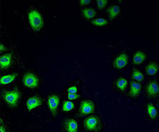
Fig2: ICC staining PDI in HUVEC cells (green). The nuclear counter stain is DAPI (blue). Cells were fixed in paraformaldehyde, permeabilised with 0.25% Triton X100/PBS.Application
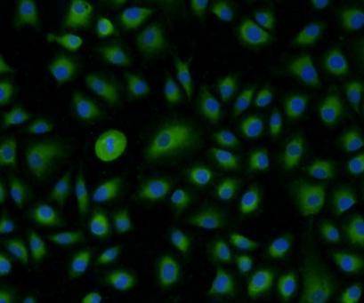
Fig3: ICC staining PDI in A549 cells (green). The nuclear counter stain is DAPI (blue). Cells were fixed in paraformaldehyde, permeabilised with 0.25% Triton X100/PBS.Application
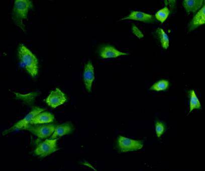
Fig4: ICC staining PDI in NIH/3T3 cells (green). The nuclear counter stain is DAPI (blue). Cells were fixed in paraformaldehyde, permeabilised with 0.25% Triton X100/PBS.Application
Fig5: Immunohistochemical analysis of paraffin-embedded human lung carcinoma tissue using anti-PDI antibody. Counter stained with hematoxylin.Application
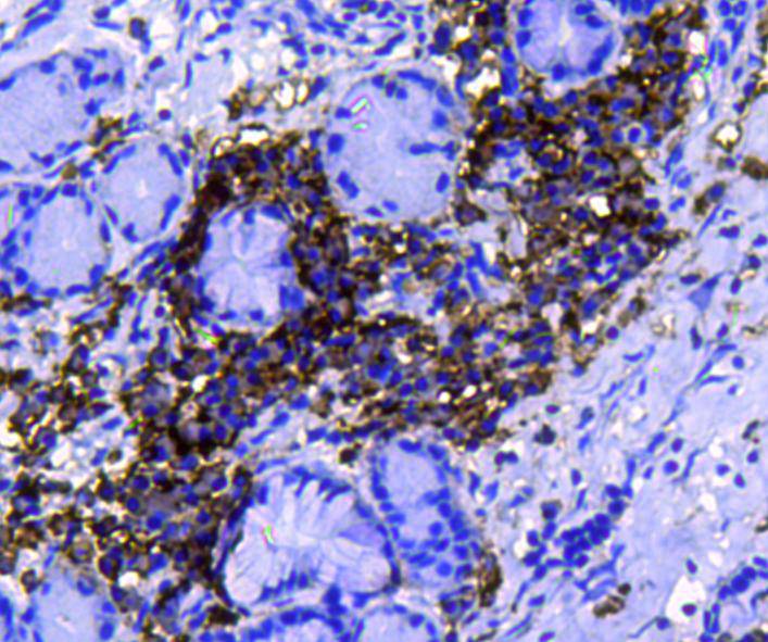
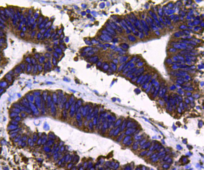
Fig6: Immunohistochemical analysis of paraffin-embedded human colon carcinoma tissue using anti-PDI antibody. Counter stained with hematoxylin.| Product Name | PDI |
|---|---|
| Antibody Type | Primary Antibodies |
| Antigen Alias | Pancreas specific protein disulfide isomerase antibody Pancreas-specific protein disulfide isomerase antibody Pancreatic protein disulfide isomerase antibody PDA2 antibody PDI antibody PDIA2 antibody PDIA2_HUMAN antibody PDIp antibody PDIR antibody Protein disulfide isomerase A2 antibody Protein disulfide isomerase antibody Protein disulfide isomerase associated 2 antibody Protein disulfide isomerase family A member 2 antibody Protein disulfide isomerase pancreatic antibody Protein disulfide-isomerase A2 antibody Rho GDP dissociation inhibitor gamma antibody |
| Product description | Protein disulfide isomerase or PDI is an enzyme in the endoplasmic reticulum in eukaryotes that catalyzes the formation and breakage of disulfide bonds between cysteine residues within proteins as they fold. Another major function of PDI relates to its activity as a chaperone; i.e., it aids wrongly folded proteins to reach a correctly folded state without the aid of enzymatic disulfide shuffling. PDI has been found to be involved in the breaking of bonds on the HIV gp120 protein during HIV infection of CD4 positive cells, and is required for HIV infection of lymphocytes and monocytes. |
| Immunogen | peptide |
| Clonality | Polyclonal |
|---|---|
| Isotype | IgG |
| Host Species | Rabbit |
| Tested Applications | WBICCIHC |
| WB:1:500-1:1,000 ICC:1:200 IHC:1:200 |
|
| Species Reactivity | HumanMouseRat |
| Concentration | 1mg/ml |
| Alternative Names | Pancreas specific protein disulfide isomerase antibody Pancreas-specific protein disulfide isomerase antibody Pancreatic protein disulfide isomerase antibody PDA2 antibody PDI antibody PDIA2 antibody PDIA2_HUMAN antibody PDIp antibody PDIR antibody Protein disulfide isomerase A2 antibody Protein disulfide isomerase antibody Protein disulfide isomerase associated 2 antibody Protein disulfide isomerase family A member 2 antibody Protein disulfide isomerase pancreatic antibody Protein disulfide-isomerase A2 antibody Rho GDP dissociation inhibitor gamma antibody |
|---|---|
| Molecular Weight(MW) | 58 kDa |
| Cellular Localization | Endoplasmic reticulum lumen |
| SwissProt ID | Q13087 |
|---|
Application
Fig1: Western blot analysis of PDI on different cell lysates using anti- PDI antibody at 1/1000 dilution. Positive control: Lane 1: Hela Lane 2: MCF-7 Lane 3: PANC Lane 4: HepG2 Lane 5: HUVEC Lane 6: NIH/3T3 Lane 7: L929 Lane 8: F9 Lane 9: Jurkat Lane 10: A431 Lane 11: A549 Lane 12: Mouse liver Lane 13: Mouse kidney Lane 14: Mouse brain Lane 15: Human kidney Lane 16: Human brain Lane 17: Human liver
Application
Fig2: ICC staining PDI in HUVEC cells (green). The nuclear counter stain is DAPI (blue). Cells were fixed in paraformaldehyde, permeabilised with 0.25% Triton X100/PBS.
Application
Fig3: ICC staining PDI in A549 cells (green). The nuclear counter stain is DAPI (blue). Cells were fixed in paraformaldehyde, permeabilised with 0.25% Triton X100/PBS.
Application
Fig4: ICC staining PDI in NIH/3T3 cells (green). The nuclear counter stain is DAPI (blue). Cells were fixed in paraformaldehyde, permeabilised with 0.25% Triton X100/PBS.
Application
Fig5: Immunohistochemical analysis of paraffin-embedded human lung carcinoma tissue using anti-PDI antibody. Counter stained with hematoxylin.
Application
Fig6: Immunohistochemical analysis of paraffin-embedded human colon carcinoma tissue using anti-PDI antibody. Counter stained with hematoxylin.| Positive Control | Hela, MCF-7, PANC,HepG2, HUVEC, NIH/3T3, L929, F9, Jurkat, A431, A549, mouse liver tissue, mouse kidney tissue, mouse brain tissue,human lung carcinoma tissue,human kidney,Human brain,Human liver,human colon carcinoma tissue. |
|---|---|
| Application Notes | WB:1:500-1:1,000 ICC:1:200 IHC:1:200 |
| Form | Liquid |
|---|---|
| Storage Instructions | Store at +4℃ after thawing. Aliquot store at -20℃ or -80℃. Avoid repeated freeze / thaw cycles. |
| Storage Buffer | 1*TBS (pH7.4), 1%BSA, 40%Glycerol. Preservative: 0.05% Sodium Azide. |


