Application
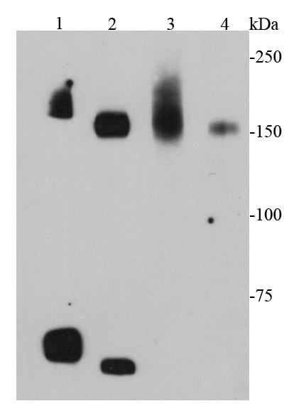
Fig1: Western blot analysis of cMet on different cell lysates using anti-cMet antibody at 1/1000 dilution.Positive control:Lane 1: Mouse liverLane 2: Mouse kidneyLane 3: D3Lane 4: MEFApplication
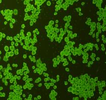
Fig2: ICC staining cMet in N2A cells (green). Cells were fixed in paraformaldehyde, permeabilised with 0.25% Triton X100/PBS.Application
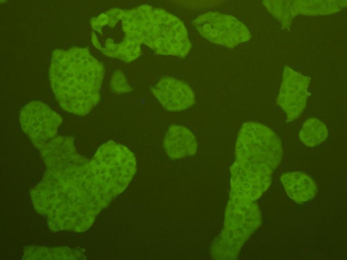
Fig3: ICC staining cMet in Hela cells (green). Cells were fixed in paraformaldehyde, permeabilised with 0.25% Triton X100/PBS.Application
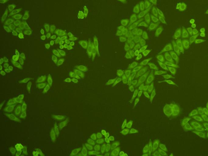
Fig4: ICC staining cMet in HepG2 cells (green). Cells were fixed in paraformaldehyde, permeabilised with 0.25% Triton X100/PBS.Application
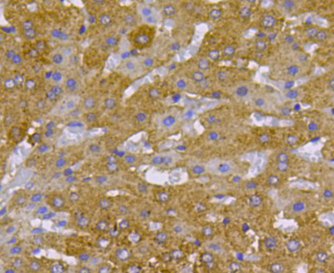
Fig5: Immunohistochemical analysis of paraffin-embedded mouse liver tissue using anti-cMet antibody. Counter stained with hematoxylin.Application
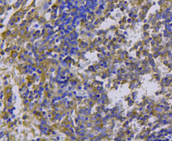
Fig6: Immunohistochemical analysis of paraffin-embedded mouse spleen tissue using anti-cMet antibody. Counter stained with hematoxylin.Application
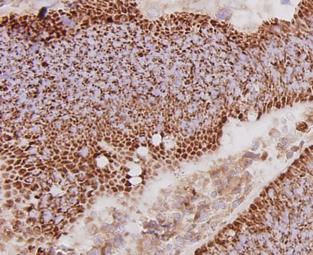
Fig7: Immunohistochemical analysis of paraffin-embedded human colon cancer tissue using anti-cMet antibody. Counter stained with hematoxylin.Application
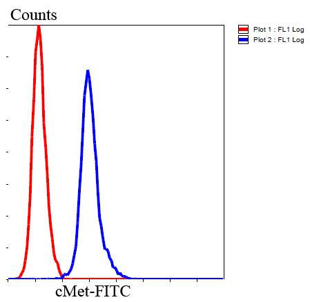
Fig8: Flow cytometric analysis of Hela cells with cMet antibody at 1/100 dilution (blue) compared with an unlabelled control (cells without incubation with primary antibody; red). Goat anti rabbit IgG (FITC) was used as the secondary antibody.| Product Name | cMet |
|---|---|
| Antibody Type | Primary Antibodies |
| Antigen Alias | AUTS9 antibody c met antibody D249 antibody Hepatocyte growth factor receptor antibody HGF antibody HGF receptor antibody HGF/SF receptor antibody HGFR antibody MET antibody Met proto oncogene tyrosine kinase antibody MET proto oncogene, receptor tyrosine kinase antibody Met proto-oncogene (hepatocyte growth factor receptor) antibody Met proto-oncogene antibody Met protooncogene antibody MET_HUMAN antibody Oncogene MET antibody Par4 antibody Proto-oncogene c-Met antibody RCCP2 antibody Scatter factor receptor antibody SF receptor antibody Tyrosine-protein kinase Met antibody |
| Product description | c-Met (MET or MNNG HOS Transforming gene) is a proto-oncogene that encodes a protein known as hepatocyte growth factor receptor (HGFR). MET is a membrane receptor that is essential for embryonic development and wound healing. Hepatocyte growth factor (HGF) is the only known ligand of the MET receptor. MET is normally expressed by cells of epithelial origin, while expression of HGF is restricted to cells of mesenchymal origin. Upon HGF stimulation, MET induces several biological responses that collectively give rise to a program known as invasive growth. Abnormal MET activation in cancer correlates with poor prognosis, where aberrantly active MET triggers tumor growth, formation of new blood vessels (angiogenesis) that supply the tumor with nutrients, and cancer spread to other organs (metastasis). MET is deregulated in many types of human malignancies, including cancers of kidney, liver, stomach, breast, and brain. |
| Immunogen | peptide |
| Clonality | Polyclonal |
|---|---|
| Isotype | IgG |
| Host Species | Rabbit |
| Tested Applications | WBICCIHCFC |
| WB:1:1,000 ICC:1:200 IHC:1:200 FC:1:100-1:200 |
|
| Species Reactivity | HumanMouseRat |
| Concentration | 1mg/ml |
| Alternative Names | AUTS9 antibody
c met antibody
D249 antibody
Hepatocyte growth factor receptor antibody
HGF antibody
HGF receptor antibody
HGF/SF receptor antibody
HGFR antibody
MET antibody
Met proto oncogene tyrosine kinase antibody
MET proto oncogene receptor tyrosine kinase antibody Met proto-oncogene (hepatocyte growth factor receptor) antibody Met proto-oncogene antibody Met protooncogene antibody MET_HUMAN antibody Oncogene MET antibody Par4 antibody Proto-oncogene c-Met antibody RCCP2 antibody Scatter factor receptor antibody SF receptor antibody Tyrosine-protein kinase Met antibody |
|---|---|
| Molecular Weight(MW) | 153 kDa |
| Cellular Localization | Cell membrane. |
| SwissProt ID | P16056 |
|---|
Application
Fig1: Western blot analysis of cMet on different cell lysates using anti-cMet antibody at 1/1000 dilution.Positive control:Lane 1: Mouse liverLane 2: Mouse kidneyLane 3: D3Lane 4: MEF
Application
Fig2: ICC staining cMet in N2A cells (green). Cells were fixed in paraformaldehyde, permeabilised with 0.25% Triton X100/PBS.
Application
Fig3: ICC staining cMet in Hela cells (green). Cells were fixed in paraformaldehyde, permeabilised with 0.25% Triton X100/PBS.
Application
Fig4: ICC staining cMet in HepG2 cells (green). Cells were fixed in paraformaldehyde, permeabilised with 0.25% Triton X100/PBS.
Application
Fig5: Immunohistochemical analysis of paraffin-embedded mouse liver tissue using anti-cMet antibody. Counter stained with hematoxylin.
Application
Fig6: Immunohistochemical analysis of paraffin-embedded mouse spleen tissue using anti-cMet antibody. Counter stained with hematoxylin.
Application
Fig7: Immunohistochemical analysis of paraffin-embedded human colon cancer tissue using anti-cMet antibody. Counter stained with hematoxylin.
Application
Fig8: Flow cytometric analysis of Hela cells with cMet antibody at 1/100 dilution (blue) compared with an unlabelled control (cells without incubation with primary antibody; red). Goat anti rabbit IgG (FITC) was used as the secondary antibody.| Positive Control | D3, MEF, Hela, N2A, mouse liver tissue, mouse kidney tissue, human colon cancer, rat liver tissue, rat spleen tissue. |
|---|---|
| Application Notes | WB:1:1,000 ICC:1:200 IHC:1:200 FC:1:100-1:200 |
| Form | Liquid |
|---|---|
| Storage Instructions | Store at +4℃ after thawing. Aliquot store at -20℃ or -80℃. Avoid repeated freeze / thaw cycles. |
| Storage Buffer | 1*TBS (pH7.4), 1%BSA, 40%Glycerol. Preservative: 0.05% Sodium Azide. |


