Application
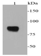
Fig1: Western blot analysis of chromogranin A on PC12 cell lysates using anti-chromogranin A antibody at 1/2000 dilution.Application
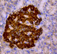
Fig2: Immunohistochemical analysis of paraffin-embedded rat pancreas tissue using anti-chromogranin A antibody. Counter stained with hematoxylin.Application
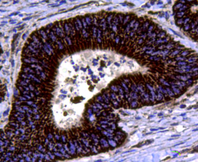
Fig3: Immunohistochemical analysis of paraffin-embedded human colon cancer tissue using anti-chromogranin A antibody. Counter stained with hematoxylin.Application
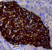
Fig4: Immunohistochemical analysis of paraffin-embedded mouse pancreas tissue using anti-chromogranin A antibody. Counter stained with hematoxylin.Application
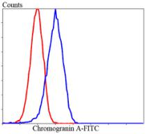
Fig5: Flow cytometric analysis of Lovo cells with chromogranin A antibody at 1/100 dilution (blue) compared with an unlabelled control (cells without incubation with primary antibody; red). Goat anti rabbit IgG (FITC) was used as the secondary antibody.| Product Name | Chromogranin A |
|---|---|
| Antibody Type | Primary Antibodies |
| Antigen Alias | CISY_HUMAN antibody Citrate synthase antibody Citrate synthase, mitochondrial antibody citrate synthetase antibody Cs antibody EC 2.3.3 antibody EC 2.3.3.1 antibody |
| Product description | Chromogranin A is a member of the chromogranin/secretogranin family of neuroendocrine secretory proteins. It is located in secretory vesicles of neurons and endocrine cells. Chromogranin A is the precursor to several functional peptides including vasostatin, pancreastatin, catestatin and parastatin. These peptides negatively modulate the neuroendocrine function of the releasing cell (autocrine) or nearby cells (paracrine). Chromogranin A might promote the generation of secretory granules. It is used as an indicator for pancreas and prostate cancer. It might play a role in early neoplasic progression. It is also elevated in diabetes. |
| Immunogen | peptide |
| Clonality | Polyclonal |
|---|---|
| Isotype | IgG |
| Host Species | Rabbit |
| Tested Applications | WBIHCFC |
| WB:1:2,000 IHC:1:200 FC:1:100-1:200 |
|
| Species Reactivity | HumanMouseRat |
| Concentration | 1mg/ml |
| Alternative Names | CISY_HUMAN antibody
Citrate synthase antibody
Citrate synthase mitochondrial antibody citrate synthetase antibody Cs antibody EC 2.3.3 antibody EC 2.3.3.1 antibody |
|---|---|
| Molecular Weight(MW) | 86 kDa |
| Cellular Localization | Cytoplasmic vesicle, secreted. |
| SwissProt ID | P26339 |
|---|
Application
Fig1: Western blot analysis of chromogranin A on PC12 cell lysates using anti-chromogranin A antibody at 1/2000 dilution.
Application
Fig2: Immunohistochemical analysis of paraffin-embedded rat pancreas tissue using anti-chromogranin A antibody. Counter stained with hematoxylin.
Application
Fig3: Immunohistochemical analysis of paraffin-embedded human colon cancer tissue using anti-chromogranin A antibody. Counter stained with hematoxylin.
Application
Fig4: Immunohistochemical analysis of paraffin-embedded mouse pancreas tissue using anti-chromogranin A antibody. Counter stained with hematoxylin.
Application
Fig5: Flow cytometric analysis of Lovo cells with chromogranin A antibody at 1/100 dilution (blue) compared with an unlabelled control (cells without incubation with primary antibody; red). Goat anti rabbit IgG (FITC) was used as the secondary antibody.| Positive Control | PC12, Lovo, mouse pancreas tissue, rat pancreas tissue, human colon cancer tissue. |
|---|---|
| Application Notes | WB:1:2,000 IHC:1:200 FC:1:100-1:200 |
| Form | Liquid |
|---|---|
| Storage Instructions | Store at +4℃ after thawing. Aliquot store at -20℃ or -80℃. Avoid repeated freeze / thaw cycles. |
| Storage Buffer | 1*TBS (pH7.4), 1%BSA, 40%Glycerol. Preservative: 0.05% Sodium Azide. |


