Application
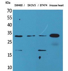
Fig1: Western blot analysis of FGF18 on human small intestine tissue lysate. Proteins were transferred to a PVDF membrane and blocked with 5% BSA in PBS for 1 hour at room temperature. The primary antibody (ER60001, 1/500) was used in 5% BSA at room temperature for 2 hours. Goat Anti-Rabbit IgG - HRP Secondary Antibody (HA1001) at 1:5,000 dilution was used for 1 hour at room temperature. Positive control: Lane 1: SW480 cell lysates Lane 2: SKOV3 cell lysates Lane 3: BT474 cell lysates Lane 4: Mouse heart tissue lysatesApplication
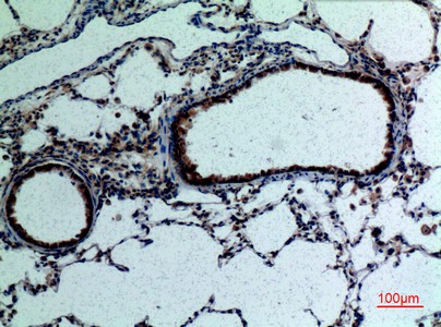
Fig2: Immunohistochemical analysis of paraffin-embedded rat lung tissue using anti-FGF18 antibody. The section was pre-treated using heat mediated antigen retrieval with Tris-EDTA buffer (pH 8.0-8.4) for 20 minutes.The tissues were blocked in 5% BSA for 30 minutes at room temperature, washed with ddH2O and PBS, and then probed with the primary antibody (ER60001, 1/200) for 30 minutes at room temperature. The detection was performed using an HRP conjugated compact polymer system. DAB was used as the chromogen. Tissues were counterstained with hematoxylin and mounted with DPX.| Product Name | FGF-18 |
|---|---|
| Antibody Type | Primary Antibodies |
| Antigen Alias | FGF 18 antibody FGF-18 antibody Fgf18 antibody FGF18_HUMAN antibody FGFI antibody Fibroblast growth factor 18 antibody zFGF5 antibody |
| Product description | Fibroblast growth factor 18 is a protein that in humans is encoded by the FGF18 gene. The protein encoded by this gene is a member of the fibroblast growth factor (FGF) family. FGF family members possess broad mitogenic and cell survival activities, and are involved in a variety of biological processes, including embryonic development, cell growth, morphogenesis, tissue repair, tumor growth, and invasion. It has been shown in vitro that this protein is able to induce neurite outgrowth in PC12 cells. FGF18 signals through fibroblast growth factor receptor FGFR3 to promote chondrogenesis and has been shown to cause thickening of cartilage in a murine model of osteoarthritis, and the recombinant version of it (sprifermin) will soon enter trials as a potential treatment for osteoarthritis. Studies of the similar proteins in mouse and chick suggested that this protein is a pleiotropic growth factor that stimulates proliferation in a number of tissues, most notably the liver and small intestine. Knockout studies of the similar gene in mice implied the role of this protein in regulating proliferation and differentiation of midline cerebellar structures. |
| Immunogen | The antiserum was produced against synthesized peptide derived from the C-terminal region of human FGF18. AA range:158-207. |
| Clonality | Polyclonal |
|---|---|
| Isotype | IgG |
| Host Species | Rabbit |
| Tested Applications | WBIHC-P |
| WB:1:500-2,000 IHC-P:1:50-1:200 |
|
| Species Reactivity | HumanMouseRat |
| Concentration | 1mg/ml |
| Alternative Names | FGF 18 antibody
FGF-18 antibody
Fgf18 antibody FGF18_HUMAN antibody FGFI antibody Fibroblast growth factor 18 antibody zFGF5 antibody |
|---|---|
| Molecular Weight(MW) | 28 kDa |
| Cellular Localization | Secreted. |
| SwissProt ID | O76093 |
|---|
Application
Fig1: Western blot analysis of FGF18 on human small intestine tissue lysate. Proteins were transferred to a PVDF membrane and blocked with 5% BSA in PBS for 1 hour at room temperature. The primary antibody (ER60001, 1/500) was used in 5% BSA at room temperature for 2 hours. Goat Anti-Rabbit IgG - HRP Secondary Antibody (HA1001) at 1:5,000 dilution was used for 1 hour at room temperature. Positive control: Lane 1: SW480 cell lysates Lane 2: SKOV3 cell lysates Lane 3: BT474 cell lysates Lane 4: Mouse heart tissue lysates
Application
Fig2: Immunohistochemical analysis of paraffin-embedded rat lung tissue using anti-FGF18 antibody. The section was pre-treated using heat mediated antigen retrieval with Tris-EDTA buffer (pH 8.0-8.4) for 20 minutes.The tissues were blocked in 5% BSA for 30 minutes at room temperature, washed with ddH2O and PBS, and then probed with the primary antibody (ER60001, 1/200) for 30 minutes at room temperature. The detection was performed using an HRP conjugated compact polymer system. DAB was used as the chromogen. Tissues were counterstained with hematoxylin and mounted with DPX.| Positive Control | SW480 cell, SKOV3 cell, BT474 cell, mouse heart, rat-lung tissue. |
|---|---|
| Application Notes | WB:1:500-2,000 IHC-P:1:50-1:200 |
| Form | Liquid |
|---|---|
| Storage Instructions | Store at +4℃ after thawing. Aliquot store at -20℃. Avoid repeated freeze / thaw cycles. |
| Storage Buffer | Liquid in PBS containing 50% glycerol, 0.5% BSA and 0.02% sodium azide. |


