Application
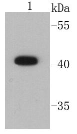
Fig1: Western blot analysis of CD9 on HepG2 cell lysates using anti-CD9 antibody at 1/1000 dilution.Application
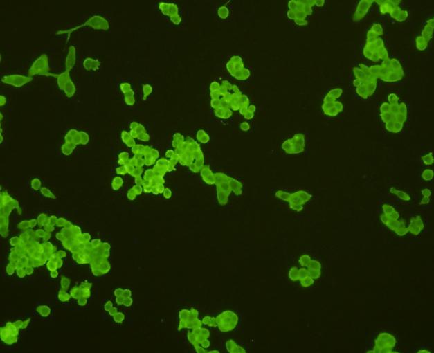
Fig2: ICC staining CD9 in Lovo cells (green). Cells were fixed in paraformaldehyde, permeabilised with 0.25% Triton X100/PBS.Application
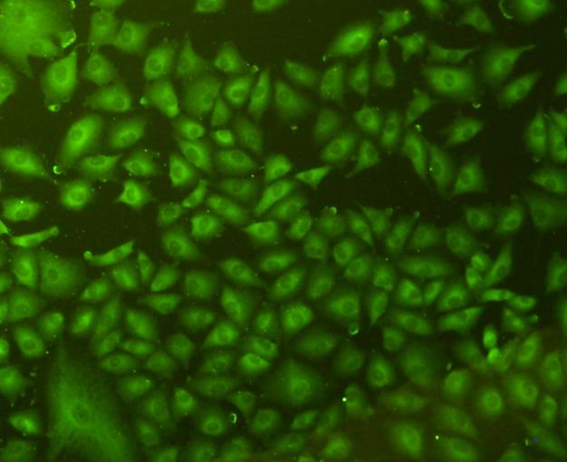
Fig3: ICC staining CD9 in A549 cells (green). Cells were fixed in paraformaldehyde, permeabilised with 0.25% Triton X100/PBS.Application
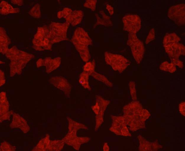
Fig4: ICC staining CD9 in Hela cells (red). Cells were fixed in paraformaldehyde, permeabilised with 0.25% Triton X100/PBS.Application
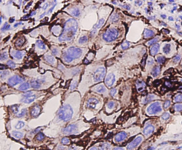
Fig5: Immunohistochemical analysis of paraffin-embedded human lung cancer tissue using anti-CD9 antibody. Counter stained with hematoxylin.Application
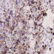
Fig6: Immunohistochemical analysis of paraffin-embedded human colon cancer tissue using anti-CD9 antibody. Counter stained with hematoxylin.Application
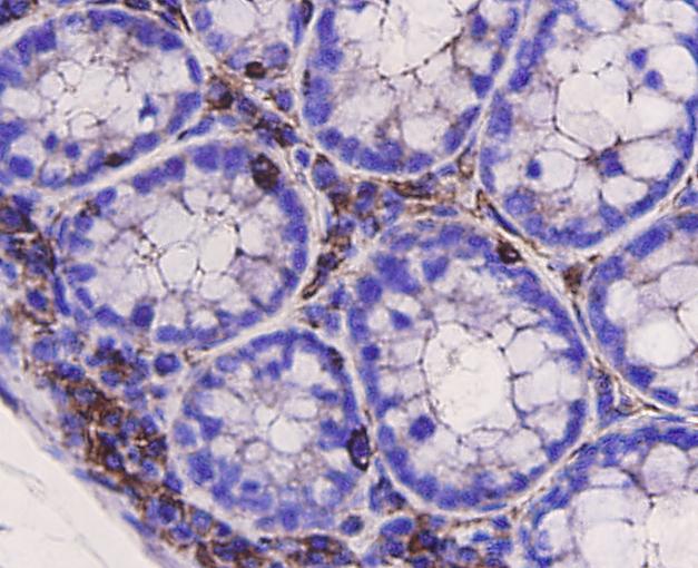
Fig7: Immunohistochemical analysis of paraffin-embedded mouse large intestine tissue using anti-CD9 antibody. Counter stained with hematoxylin.Application
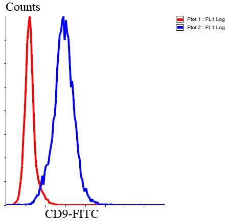
Fig8: Flow cytometric analysis of Jurkat cells with CD9 antibody at 1/100 dilution (blue) compared with an unlabelled control (cells without incubation with primary antibody; red). Goat anti rabbit IgG (FITC) was used as the secondary antibody.| Product Name | CD9 |
|---|---|
| Antibody Type | Primary Antibodies |
| Antigen Alias | Tetraspanin 29 antibody 5H9 antibody 5H9 antigen antibody Antigen defined by monoclonal antibody 602 29 antibody Antigen defined by monoclonal antibody 60229 antibody BA-2/p24 antigen antibody BA2 antibody BTCC 1 antibody BTCC1 antibody CD9 antibody CD9 antigen antibody CD9 antigen p24 antibody CD9 molecule antibody CD9_HUMAN antibody Cell growth inhibiting gene 2 protein antibody Cell growth-inhibiting gene 2 protein antibody DRAP 27 antibody DRAP27 antibody GIG2 antibody Growth inhibiting gene 2 protein antibody Leukocyte antigen MIC3 antibody MIC3 antibody Motility related protein antibody Motility-related protein antibody MRP 1 antibody MRP-1 antibody MRP1 antibody p24 antibody p24 antigen antibody Tetraspanin-29 antibody Tspan 29 antibody Tspan-29 antibody TSPAN29 antibody |
| Product description | CD9 antigen is a glycoprotein expressed on the surface of developing B lymphocytes, platelets, monocytes, eosinophils, basophil, stimulated T lymphocytes and by neurons and glial cells in the peripheral nervous system. Protein exists in three forms, and is known to carry covalently linked fatty acids. It is involved in platelet activation, aggregation, in cell adhesion, cell motility and tumor metastasis. It regulates paranodal junction formation, and is also required for gamete fusion. |
| Immunogen | peptide |
| Clonality | Polyclonal |
|---|---|
| Isotype | IgG |
| Host Species | Rabbit |
| Tested Applications | WBICCIHCFC |
| WB:1:1,000 ICC:1:200 IHC:1:200 FC:1:100-1:200 |
|
| Species Reactivity | HumanMouseRat |
| Concentration | 1mg/ml |
| Alternative Names | Tetraspanin 29 antibody 5H9 antibody 5H9 antigen antibody Antigen defined by monoclonal antibody 602 29 antibody Antigen defined by monoclonal antibody 60229 antibody BA-2/p24 antigen antibody BA2 antibody BTCC 1 antibody BTCC1 antibody CD9 antibody CD9 antigen antibody CD9 antigen p24 antibody CD9 molecule antibody CD9_HUMAN antibody Cell growth inhibiting gene 2 protein antibody Cell growth-inhibiting gene 2 protein antibody DRAP 27 antibody DRAP27 antibody GIG2 antibody Growth inhibiting gene 2 protein antibody Leukocyte antigen MIC3 antibody MIC3 antibody Motility related protein antibody Motility-related protein antibody MRP 1 antibody MRP-1 antibody MRP1 antibody p24 antibody p24 antigen antibody Tetraspanin-29 antibody Tspan 29 antibody Tspan-29 antibody TSPAN29 antibody |
|---|---|
| Molecular Weight(MW) | 25 kDa |
| Cellular Localization | Cell membrane. |
| SwissProt ID | P21926 |
|---|
Application
Fig1: Western blot analysis of CD9 on HepG2 cell lysates using anti-CD9 antibody at 1/1000 dilution.
Application
Fig2: ICC staining CD9 in Lovo cells (green). Cells were fixed in paraformaldehyde, permeabilised with 0.25% Triton X100/PBS.
Application
Fig3: ICC staining CD9 in A549 cells (green). Cells were fixed in paraformaldehyde, permeabilised with 0.25% Triton X100/PBS.
Application
Fig4: ICC staining CD9 in Hela cells (red). Cells were fixed in paraformaldehyde, permeabilised with 0.25% Triton X100/PBS.
Application
Fig5: Immunohistochemical analysis of paraffin-embedded human lung cancer tissue using anti-CD9 antibody. Counter stained with hematoxylin.
Application
Fig6: Immunohistochemical analysis of paraffin-embedded human colon cancer tissue using anti-CD9 antibody. Counter stained with hematoxylin.
Application
Fig7: Immunohistochemical analysis of paraffin-embedded mouse large intestine tissue using anti-CD9 antibody. Counter stained with hematoxylin.
Application
Fig8: Flow cytometric analysis of Jurkat cells with CD9 antibody at 1/100 dilution (blue) compared with an unlabelled control (cells without incubation with primary antibody; red). Goat anti rabbit IgG (FITC) was used as the secondary antibody.| Positive Control | A549, Hela, Jurkat, HepG2, Lovo, human lung cancer tissue, human colon cancer tissue, mouse large intestine tissue. |
|---|---|
| Application Notes | WB:1:1,000 ICC:1:200 IHC:1:200 FC:1:100-1:200 |
| Form | Liquid |
|---|---|
| Storage Instructions | Store at +4℃ after thawing. Aliquot store at -20℃ or -80℃. Avoid repeated freeze / thaw cycles. |
| Storage Buffer | 1*TBS (pH7.4), 1%BSA, 40%Glycerol. Preservative: 0.05% Sodium Azide. |


