Application
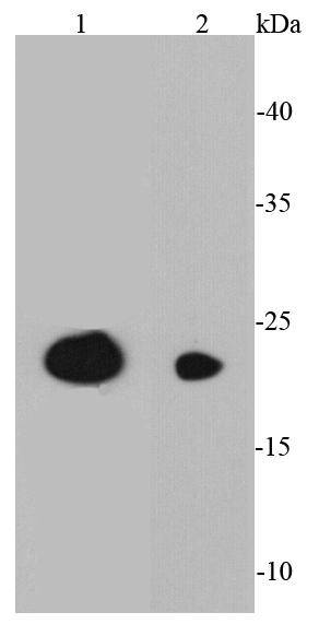
Fig1: Western blot analysis of CD3E on different cell lysates using anti-CD3E antibody at 1/1000 dilution.Application
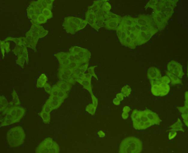
Fig2: ICC staining CD3E in Hela cells (green). Cells were fixed in paraformaldehyde, permeabilised with 0.25% Triton X100/PBS.Application
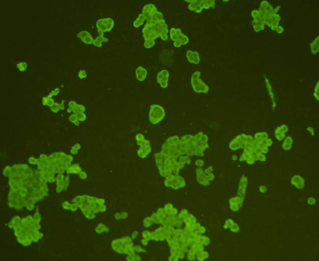
Fig3: ICC staining CD3E in Lovo cells (green). Cells were fixed in paraformaldehyde, permeabilised with 0.25% Triton X100/PBS.Application
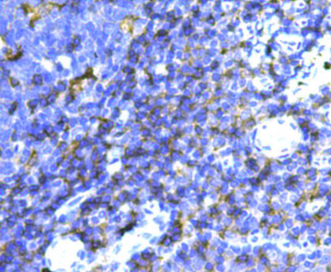
Fig4: Immunohistochemical analysis of paraffin-embedded human tonsil tissue using anti-CD3E antibody. Counter stained with hematoxylin.Application
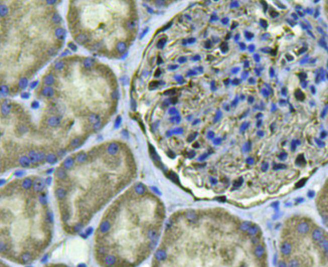
Fig5: Immunohistochemical analysis of paraffin-embedded human kidney tissue using anti-CD3E antibody. Counter stained with hematoxylin.Application
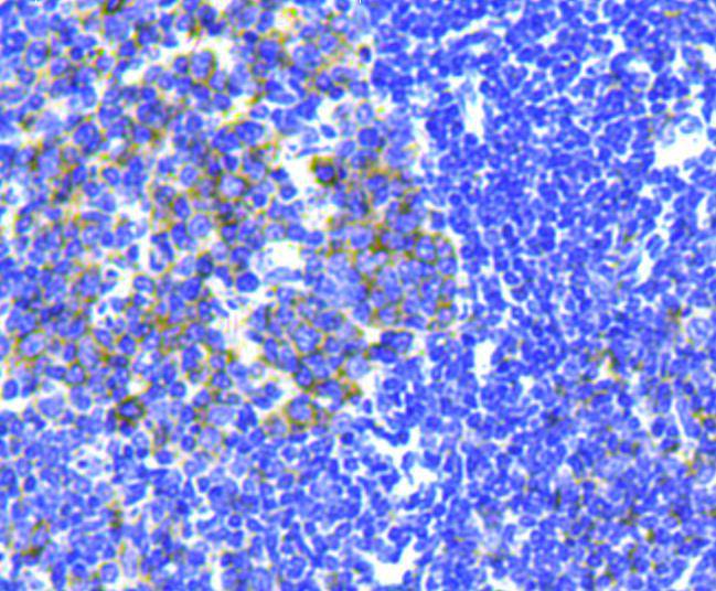
Fig6: Immunohistochemical analysis of paraffin-embedded mouse spleen tissue using anti-CD3E antibody. Counter stained with hematoxylin.Application
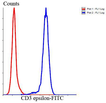
Fig7: Flow cytometric analysis of Jurkat cells with CD3E antibody at 1/100 dilution (blue) compared with an unlabelled control (cells without incubation with primary antibody; red). Goat anti rabbit IgG (FITC) was used as the secondary antibody.| Product Name | CD3 epsilon |
|---|---|
| Antibody Type | Primary Antibodies |
| Antigen Alias | CD3 epsilon antibody CD3e antibody CD3e antigen epsilon polypeptide (TiT3 complex) antibody CD3E antigen epsilon polypeptide antibody CD3E antigen, epsilon subunit antibody CD3e molecule epsilon antibody CD3e molecule, epsilon (CD3 TCR complex) antibody CD3e molecule, epsilon (CD3-TCR complex) antibody CD3E_HUMAN antibody IMD18 antibody T cell antigen receptor complex epsilon subunit of T3 antibody T cell surface antigen T3/Leu 4 epsilon chain antibody T cell surface glycoprotein CD3 epsilon chain antibody T-cell surface antigen T3/Leu-4 epsilon chain antibody T-cell surface glycoprotein CD3 epsilon chain antibody T3E antibody TCRE antibody |
| Product description | CD3 is composed of five invariant polypeptide chains that associate to form three dimers. The five invariant chains of CD3 are labeled gamma, delta, epsilon, zeta, and eta. Each of these polypeptides contains at least one immunoreceptor tyrosine-based activation motif (ITAM) These chains associate with the T cell receptor (TCR) and the ζ-chain to generate an activation signal in T lymphocytes. |
| Immunogen | This antibody is produced by immunizing rabbits with a synthetic peptide (KLH-coupled) corresponding to N-terminal residues of CD3 epsilon. |
| Clonality | Polyclonal |
|---|---|
| Isotype | IgG |
| Host Species | Rabbit |
| Tested Applications | WBICCIHCFC |
| WB:1:1,000 ICC:1:200 IHC:1:200 FC:1:100-1:200 |
|
| Species Reactivity | HumanMouse |
| Concentration | 1mg/ml |
| Alternative Names | CD3 epsilon antibody
CD3e antibody
CD3e antigen epsilon polypeptide (TiT3 complex) antibody
CD3E antigen epsilon polypeptide antibody
CD3E antigen epsilon subunit antibody CD3e molecule epsilon antibody CD3e molecule epsilon (CD3 TCR complex) antibody CD3e molecule epsilon (CD3-TCR complex) antibody CD3E_HUMAN antibody IMD18 antibody T cell antigen receptor complex epsilon subunit of T3 antibody T cell surface antigen T3/Leu 4 epsilon chain antibody T cell surface glycoprotein CD3 epsilon chain antibody T-cell surface antigen T3/Leu-4 epsilon chain antibody T-cell surface glycoprotein CD3 epsilon chain antibody T3E antibody TCRE antibody |
|---|---|
| Molecular Weight(MW) | 23 kDa |
| Cellular Localization | Membrane. |
Application
Fig1: Western blot analysis of CD3E on different cell lysates using anti-CD3E antibody at 1/1000 dilution.
Application
Fig2: ICC staining CD3E in Hela cells (green). Cells were fixed in paraformaldehyde, permeabilised with 0.25% Triton X100/PBS.
Application
Fig3: ICC staining CD3E in Lovo cells (green). Cells were fixed in paraformaldehyde, permeabilised with 0.25% Triton X100/PBS.
Application
Fig4: Immunohistochemical analysis of paraffin-embedded human tonsil tissue using anti-CD3E antibody. Counter stained with hematoxylin.
Application
Fig5: Immunohistochemical analysis of paraffin-embedded human kidney tissue using anti-CD3E antibody. Counter stained with hematoxylin.
Application
Fig6: Immunohistochemical analysis of paraffin-embedded mouse spleen tissue using anti-CD3E antibody. Counter stained with hematoxylin.
Application
Fig7: Flow cytometric analysis of Jurkat cells with CD3E antibody at 1/100 dilution (blue) compared with an unlabelled control (cells without incubation with primary antibody; red). Goat anti rabbit IgG (FITC) was used as the secondary antibody.| Positive Control | Hela, Jurkat, LOVO, human thymus tissue, human tonsil tissue, human kidney tissue, mouse spleen tissue |
|---|---|
| Application Notes | WB:1:1,000 ICC:1:200 IHC:1:200 FC:1:100-1:200 |
| Form | Liquid |
|---|---|
| Storage Instructions | Store at +4℃ after thawing. Aliquot store at -20℃. Avoid repeated freeze / thaw cycles. |
| Storage Buffer | 1*TBS (pH7.4), 1%BSA, 40%Glycerol. Preservative: 0.05% Sodium Azide. |


