Application
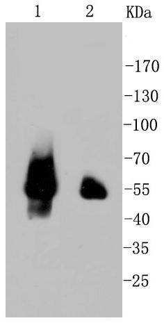
Fig1: Western blot analysis of p53 on different cell lysates using anti-p53 antibody at 1/1,000 dilution. Positive control: Lane 1: 293T Lane 2: 293Application
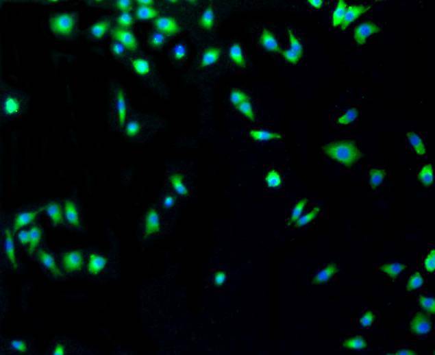
Fig2: ICC staining p53 in A549 cells (green). The nuclear counter stain is DAPI (blue). Cells were fixed in paraformaldehyde, permeabilised with 0.25% Triton X100/PBS.Application
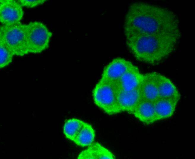
Fig3: ICC staining p53 in SW480 cells (green). The nuclear counter stain is DAPI (blue). Cells were fixed in paraformaldehyde, permeabilised with 0.25% Triton X100/PBS.Application
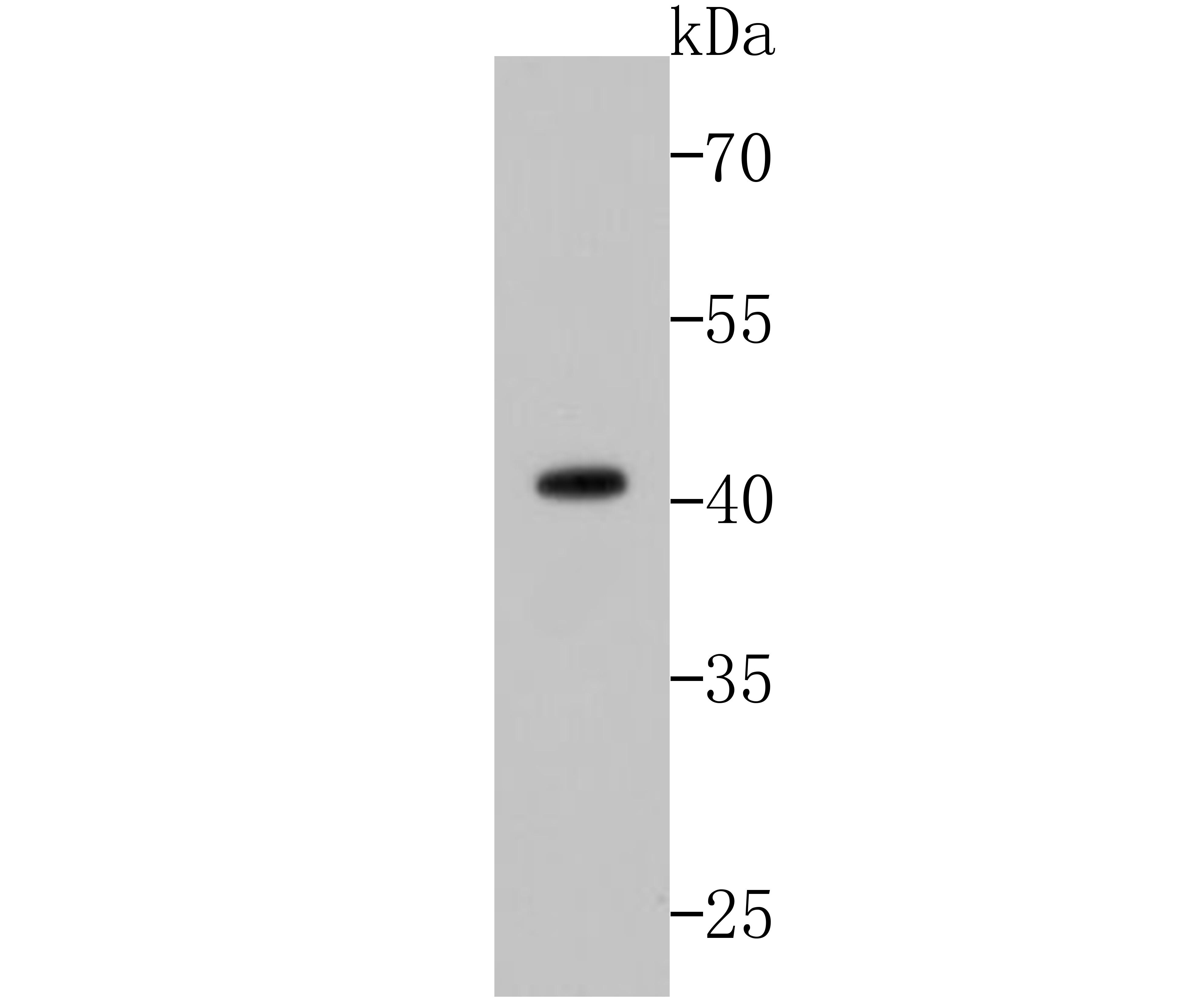
Fig4: Western blot analysis of p53 on zebrafish tissue lysates using anti-p53 antibody at 1/500 dilution.| Product Name | p53 [SA39-07] |
|---|---|
| Antibody Type | Primary Antibodies |
| Antigen Alias | Antigen NY-CO-13 antibody BCC7 antibody Cellular tumor antigen p53 antibody FLJ92943 antibody LFS1 antibody Mutant tumor protein 53 antibody p53 antibody p53 tumor suppressor antibody P53_HUMAN antibody Phosphoprotein p53 antibody Tp53 antibody Transformation related protein 53 antibody TRP53 antibody Tumor protein 53 antibody Tumor protein p53 antibody Tumor suppressor p53 antibody |
| Product description | The p53 tumor suppressor protein plays a major role in cellular response to DNA damage and other genomic aberrations. Activation of p53 can lead to either cell cycle arrest and DNA repair or apoptosis. It is involved in cell cycle regulation as a trans-activator that acts to negatively regulate cell division by controlling a set of genes required for this process. p53 is phosphorylated at multiple sites in vivo and by several different protein kinases in vitro. P53 is found in increased amounts in a wide variety of transformed cells. P53 is frequently mutated or inactivated in about 60% of cancers. |
| Immunogen | recombinant protein |
| Clonality | Monoclonal |
|---|---|
| Isotype | IgG |
| Host Species | Recombinant rabbit |
| Tested Applications | WBICC/IF |
| WB:1:1,000-1:5,000 ICC:1:50-1:200 |
|
| Species Reactivity | HumanZebra Fish |
| Concentration | 1mg/ml |
| Alternative Names | Antigen NY-CO-13 antibody BCC7 antibody Cellular tumor antigen p53 antibody FLJ92943 antibody LFS1 antibody Mutant tumor protein 53 antibody p53 antibody p53 tumor suppressor antibody P53_HUMAN antibody Phosphoprotein p53 antibody Tp53 antibody Transformation related protein 53 antibody TRP53 antibody Tumor protein 53 antibody Tumor protein p53 antibody Tumor suppressor p53 antibody |
|---|---|
| Molecular Weight(MW) | 44/53 kDa (Human), 41kDa (Zebrafish) |
| Cellular Localization | Cytoplasm, Nucleus, Endoplasmic reticulum, Mitochondrion matrix |
| SwissProt ID | P04637 |
|---|
Application
Fig1: Western blot analysis of p53 on different cell lysates using anti-p53 antibody at 1/1,000 dilution. Positive control: Lane 1: 293T Lane 2: 293
Application
Fig2: ICC staining p53 in A549 cells (green). The nuclear counter stain is DAPI (blue). Cells were fixed in paraformaldehyde, permeabilised with 0.25% Triton X100/PBS.
Application
Fig3: ICC staining p53 in SW480 cells (green). The nuclear counter stain is DAPI (blue). Cells were fixed in paraformaldehyde, permeabilised with 0.25% Triton X100/PBS.
Application
Fig4: Western blot analysis of p53 on zebrafish tissue lysates using anti-p53 antibody at 1/500 dilution.| Positive Control | A549, SW480. |
|---|---|
| Application Notes | WB:1:1,000-1:5,000 ICC:1:50-1:200 |
| Form | Liquid |
|---|---|
| Storage Instructions | Store at +4℃ after thawing. Aliquot store at -20℃ or -80℃. Avoid repeated freeze / thaw cycles. |
| Storage Buffer | 1*TBS (pH7.4), 1%BSA, 40%Glycerol. Preservative: 0.05% Sodium Azide. |


