Application
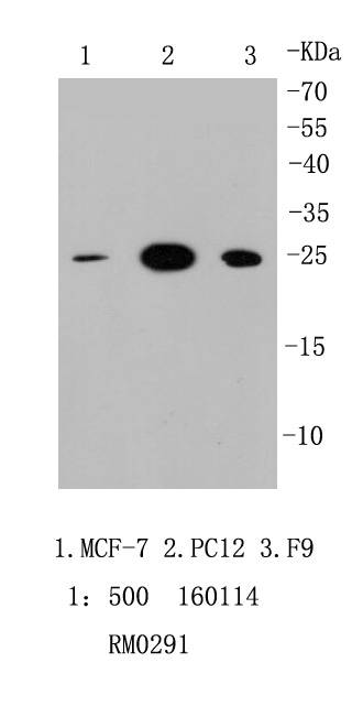
Fig1: Western blot analysis of HMGB1 on different cell lysates using anti-HMGB1 antibody at 1/1,000 dilution. Positive control: Lane 1: MCF-7 Lane 2: PC12 Lane 3: F9Application
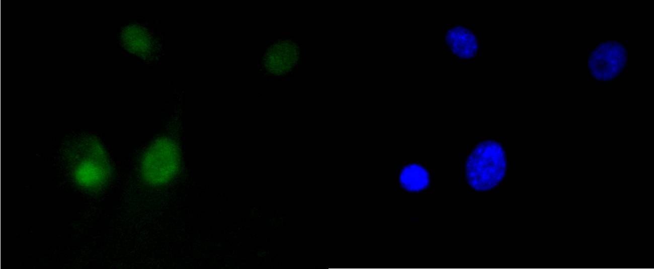
Fig2: ICC staining HMGB1 in NIH/3T3 cells (green). The nuclear counter stain is DAPI (blue). Cells were fixed in paraformaldehyde, permeabilised with 0.25% Triton X100/PBS.Application
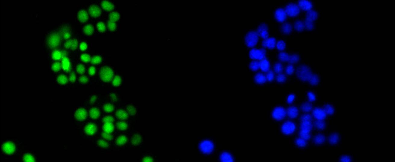
Fig3: ICC staining HMGB1 in MCF-7 cells (green). The nuclear counter stain is DAPI (blue). Cells were fixed in paraformaldehyde, permeabilised with 0.25% Triton X100/PBS.Application
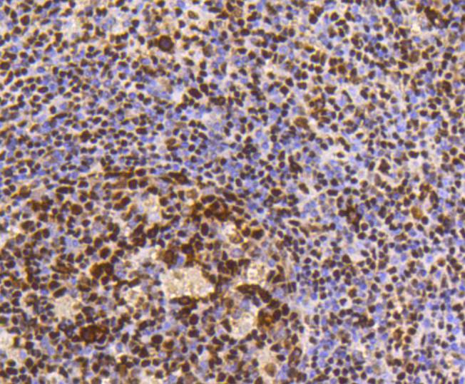
Fig4: Immunohistochemical analysis of paraffin-embedded human tonsil tissue using anti-HMGB1 antibody. Counter stained with hematoxylin.Application
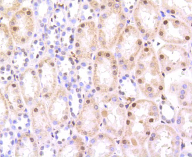
Fig5: Immunohistochemical analysis of paraffin-embedded human kidney tissue using anti-HMGB1 antibody. Counter stained with hematoxylin.Application
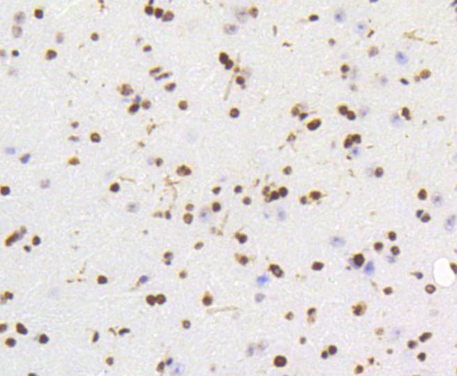
Fig6: Immunohistochemical analysis of paraffin-embedded mouse brain tissue using anti-HMGB1 antibody. Counter stained with hematoxylin.Application
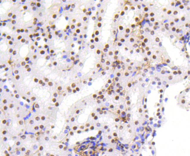
Fig7: Immunohistochemical analysis of paraffin-embedded mouse kidney tissue using anti-HMGB1 antibody. Counter stained with hematoxylin.Application
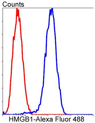
Fig8: Flow cytometric analysis of PC-12 cells with HMGB1 antibody at 1/50 dilution (blue) compared with an unlabelled control (cells without incubation with primary antibody; red). Alexa Fluor 488-conjugated goat anti-rabbit IgG was used as the secondary antibody..| Product Name | HMGB1 [SA39-03] |
|---|---|
| Antibody Type | Primary Antibodies |
| Antigen Alias | Amphoterin antibody Chromosomal protein, nonhistone, HMG1 antibody DKFZp686A04236 antibody High mobility group 1 antibody High mobility group box 1 antibody High mobility group protein 1 antibody High mobility group protein B1 antibody high-mobility group (nonhistone chromosomal) protein 1 antibody HMG-1 antibody HMG1 antibody HMG3 antibody HMGB 1 antibody HMGB1 antibody HMGB1_HUMAN antibody NONHISTONE CHROMOSOMAL PROTEIN HMG1 antibody SBP 1 antibody Sulfoglucuronyl carbohydrate binding protein antibody |
| Product description | Like the histones, HMGB1, also known as high-mobility group protein 1 (HMG-1) is among the most important chromatin proteins. In the nucleus HMGB1 interacts with nucleosomes, transcription factors, and histones. This nuclear protein organizes the DNA and regulates transcription. After binding, HMGB1 bends DNA, which facilitates the binding of other proteins. HMGB1 is secreted by immune cells (like macrophages, monocytes and dendritic cells) through leaderless secretory pathway. Activated macrophages and monocytes secrete HMGB1 as a cytokine mediator of Inflammation. In recent research, HMGB1 has been reported as a novel biomarker for human ovarian cancer |
| Immunogen | recombinant protein |
| Clonality | Monoclonal |
|---|---|
| Isotype | IgG |
| Host Species | Recombinant rabbit |
| Tested Applications | WBICC/IFIHCFC |
| WB:1:1,000-1:5,000 ICC:1:50-1:200 IHC:1:50-1:200 FC:1:10-1:100 |
|
| Species Reactivity | HumanMouseRat |
| Concentration | 1mg/ml |
| Alternative Names | Amphoterin antibody
Chromosomal protein nonhistone HMG1 antibody DKFZp686A04236 antibody High mobility group 1 antibody High mobility group box 1 antibody High mobility group protein 1 antibody High mobility group protein B1 antibody high-mobility group (nonhistone chromosomal) protein 1 antibody HMG-1 antibody HMG1 antibody HMG3 antibody HMGB 1 antibody HMGB1 antibody HMGB1_HUMAN antibody NONHISTONE CHROMOSOMAL PROTEIN HMG1 antibody SBP 1 antibody Sulfoglucuronyl carbohydrate binding protein antibody |
|---|---|
| Molecular Weight(MW) | 25 kDa |
| Cellular Localization | Cytoplasm, Nucleus, Cell membrane, Secreted, Chromosome |
Application
Fig1: Western blot analysis of HMGB1 on different cell lysates using anti-HMGB1 antibody at 1/1,000 dilution. Positive control: Lane 1: MCF-7 Lane 2: PC12 Lane 3: F9
Application
Fig2: ICC staining HMGB1 in NIH/3T3 cells (green). The nuclear counter stain is DAPI (blue). Cells were fixed in paraformaldehyde, permeabilised with 0.25% Triton X100/PBS.
Application
Fig3: ICC staining HMGB1 in MCF-7 cells (green). The nuclear counter stain is DAPI (blue). Cells were fixed in paraformaldehyde, permeabilised with 0.25% Triton X100/PBS.
Application
Fig4: Immunohistochemical analysis of paraffin-embedded human tonsil tissue using anti-HMGB1 antibody. Counter stained with hematoxylin.
Application
Fig5: Immunohistochemical analysis of paraffin-embedded human kidney tissue using anti-HMGB1 antibody. Counter stained with hematoxylin.
Application
Fig6: Immunohistochemical analysis of paraffin-embedded mouse brain tissue using anti-HMGB1 antibody. Counter stained with hematoxylin.
Application
Fig7: Immunohistochemical analysis of paraffin-embedded mouse kidney tissue using anti-HMGB1 antibody. Counter stained with hematoxylin.
Application
Fig8: Flow cytometric analysis of PC-12 cells with HMGB1 antibody at 1/50 dilution (blue) compared with an unlabelled control (cells without incubation with primary antibody; red). Alexa Fluor 488-conjugated goat anti-rabbit IgG was used as the secondary antibody..| Positive Control | NIH/3T3, MCF-7, PC12, F9, human tonsil tissue, mouse kidney tissue,mouse brain tissue, human kidney tissue. |
|---|---|
| Application Notes | WB:1:1,000-1:5,000 ICC:1:50-1:200 IHC:1:50-1:200 FC:1:10-1:100 |
| Form | Liquid |
|---|---|
| Storage Instructions | Store at +4℃ after thawing. Aliquot store at -20℃ or -80℃. Avoid repeated freeze / thaw cycles. |
| Storage Buffer | 1*TBS (pH7.4), 1%BSA, 40%Glycerol. Preservative: 0.05% Sodium Azide. |


