WB
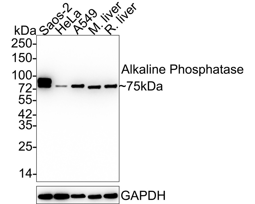
Western blot analysis of Alkaline Phosphatase on different lysates with Rabbit anti-Alkaline Phosphatase antibody at 1/5,000 dilution. Lane 1: Saos-2 cell lysate (15 µg/Lane), Lane 2: HeLa cell lysate (15 µg/Lane), Lane 3: A549 cell lysate (15 µg/Lane), Lane 4: Mouse liver tissue lysate (20 µg/Lane), Lane 5: Rat liver tissue lysate (20 µg/Lane), Exposure time: 24 seconds; 4-20% SDS-PAGE gel. Proteins were transferred to a PVDF membrane and blocked with 5% NFDM/TBST for 1 hour at room temperature. The primary antibody at 1/5,000 dilution was used in 5% NFDM/TBST at 4℃ overnight. Goat Anti-Rabbit IgG - HRP Secondary Antibody at 1:50,000 dilution was used for 1 hour at room temperature.IHC
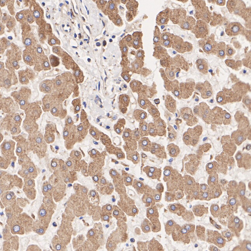
Immunohistochemical analysis of paraffin-embedded human liver tissue with Rabbit anti-Alkaline Phosphatase antibody at 1/8,000 dilution. The section was pre-treated using heat mediated antigen retrieval with Tris-EDTA buffer (pH 9.0) for 20 minutes. The tissues were blocked in 1% BSA for 20 minutes at room temperature, washed with ddH2O and PBS, and then probed with the primary antibody at 1/8,000 dilution for 1 hour at room temperature. The detection was performed using an HRP conjugated compact polymer system. DAB was used as the chromogen. Tissues were counterstained with hematoxylin and mounted with DPX.IHC
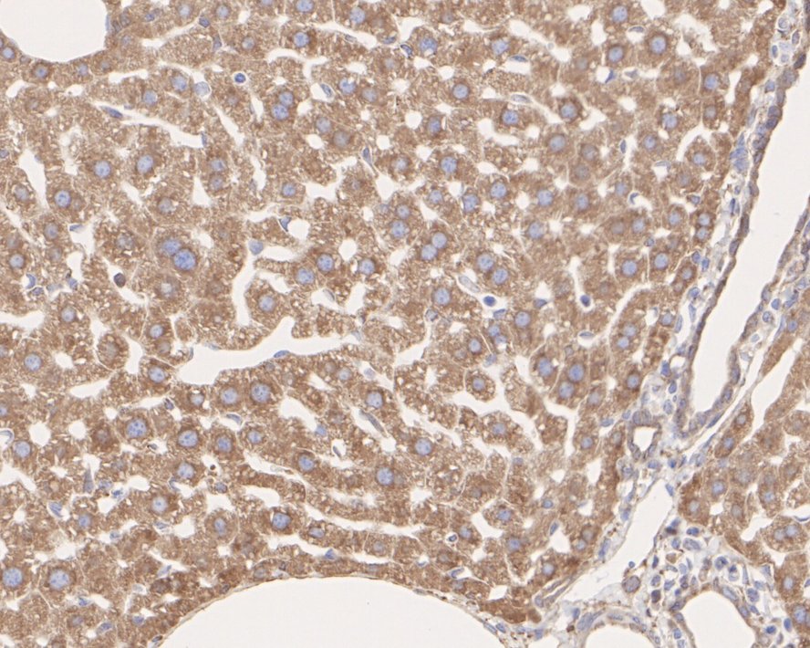
Immunohistochemical analysis of paraffin-embedded mouse liver tissue with Rabbit anti-Alkaline Phosphatase antibody at 1/8,000 dilution. The section was pre-treated using heat mediated antigen retrieval with Tris-EDTA buffer (pH 9.0) for 20 minutes. The tissues were blocked in 1% BSA for 20 minutes at room temperature, washed with ddH2O and PBS, and then probed with the primary antibody at 1/8,000 dilution for 1 hour at room temperature. The detection was performed using an HRP conjugated compact polymer system. DAB was used as the chromogen. Tissues were counterstained with hematoxylin and mounted with DPX.IHC
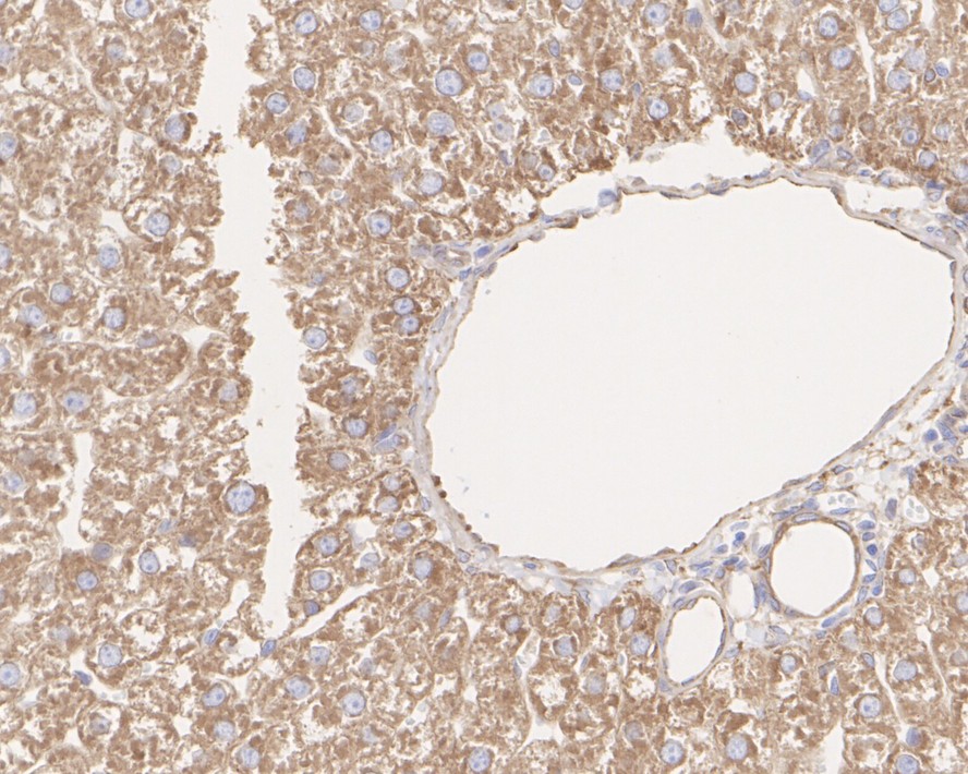
Immunohistochemical analysis of paraffin-embedded rat liver tissue with Rabbit anti-Alkaline Phosphatase antibody at 1/8,000 dilution. The section was pre-treated using heat mediated antigen retrieval with Tris-EDTA buffer (pH 9.0) for 20 minutes. The tissues were blocked in 1% BSA for 20 minutes at room temperature, washed with ddH2O and PBS, and then probed with the primary antibody at 1/8,000 dilution for 1 hour at room temperature. The detection was performed using an HRP conjugated compact polymer system. DAB was used as the chromogen. Tissues were counterstained with hematoxylin and mounted with DPX.IF-P
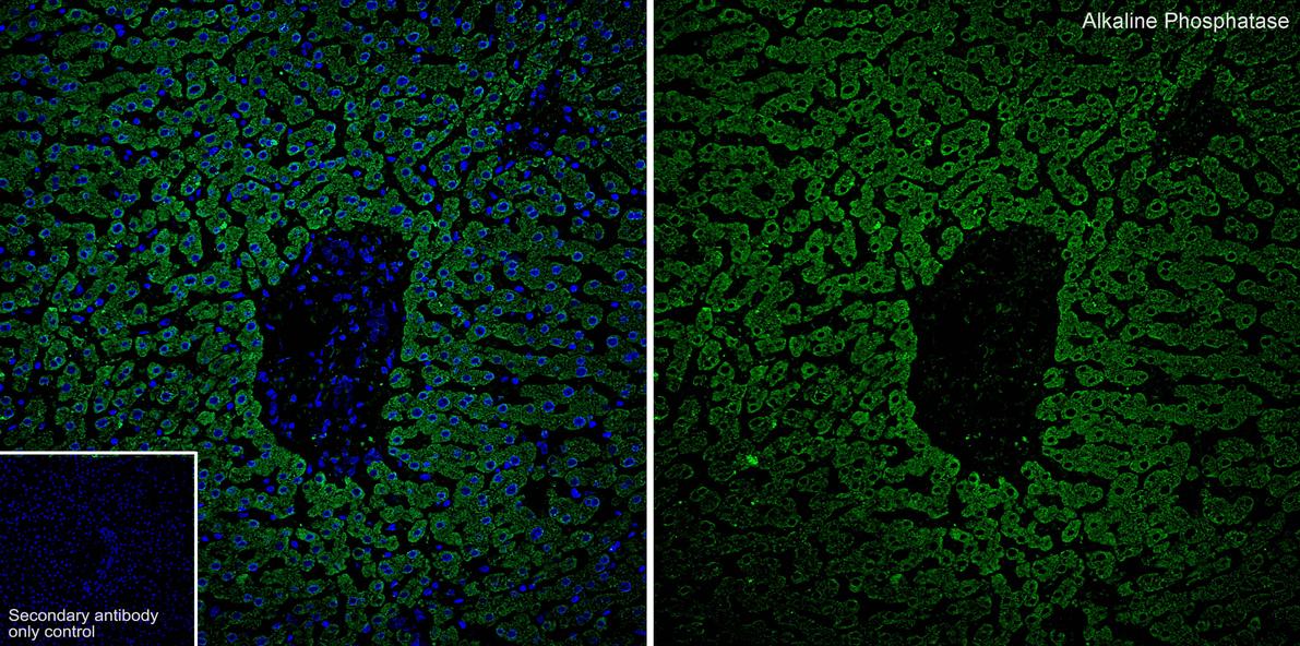
Species: Human, Site: liver, Sample: Paraffin-embedded section, Antibody concentration: 1/200.IF-P
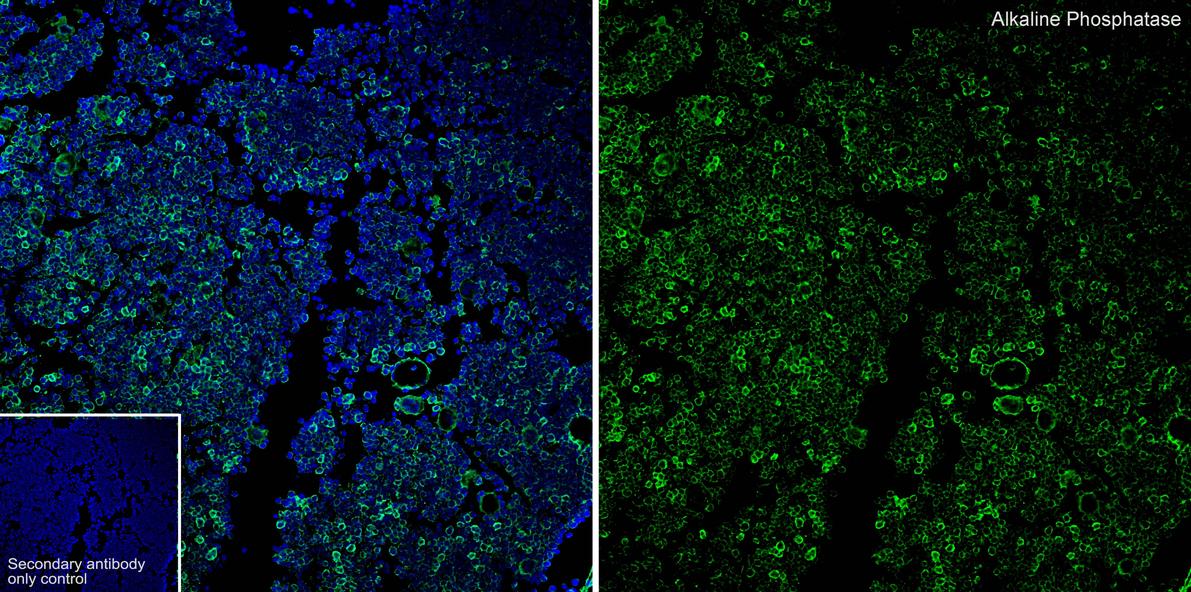
Species: Mouse, Site: bone, Sample: Paraffin-embedded section, Antibody concentration: 1/200.| Product Name | Alkaline Phosphatase Recombinant Rabbit Monoclonal Antibody |
|---|---|
| Antibody Type | Primary Antibodies |
| Product description | Alkaline phosphatase (ALP) removes phosphate groups from the 5' end of DNA and RNA, and from proteins. Most mammals have 4 different isozymes: placental, placental like, intestinal and non tissue specific (found in liver, kidney and bone). Tissues with particularly high concentrations of ALP include the liver, bile ducts, placenta, and bone. Placental ALP is highly polymorphic, there are at least three common alleles. Damaged or diseased tissue releases enzymes into the blood, so serum ALP measurements can be abnormal in many conditions, including bone disease and liver disease. |
| Immunogen | Synthetic peptide within human Alkaline Phosphatase aa 18-50. |
| Clonality | Monoclonal |
|---|---|
| Isotype | IgG |
| Host Species | Recombinant rabbit |
| Tested Applications | IF-PIHCWB |
| WB:1:5000 IHC:1:1000-1:8000 IF-P:1:200 |
|
| Species Reactivity | HumanMouseRat |
| Concentration | 1mg/ml |
| Purification | Protein A |
| Alternative Names | Alkaline phosphatase antibody Alkaline phosphatase placental antibody Alkaline phosphatase placental type antibody Alkaline phosphatase Regan isozyme antibody ALP antibody Alp1 antibody ALPP antibody FLJ61142 antibody Germ-cell alkaline phosphatase antibody nagao isozyme antibody OTTHUMP00000164354 antibody PALP antibody Placental alkaline phosphatase 1 antibody placental heat-stable alkaline phosphatase antibody placental type antibody PLAP antibody PLAP-1 antibody PLAP1 antibody PPB1_HUMAN antibody |
|---|---|
| Molecular Weight(MW) | 57 kDa(Observed band size: 75 kDa) |
| Cellular Localization | Cell membrane |
WB
Western blot analysis of Alkaline Phosphatase on different lysates with Rabbit anti-Alkaline Phosphatase antibody at 1/5,000 dilution. Lane 1: Saos-2 cell lysate (15 µg/Lane), Lane 2: HeLa cell lysate (15 µg/Lane), Lane 3: A549 cell lysate (15 µg/Lane), Lane 4: Mouse liver tissue lysate (20 µg/Lane), Lane 5: Rat liver tissue lysate (20 µg/Lane), Exposure time: 24 seconds; 4-20% SDS-PAGE gel. Proteins were transferred to a PVDF membrane and blocked with 5% NFDM/TBST for 1 hour at room temperature. The primary antibody at 1/5,000 dilution was used in 5% NFDM/TBST at 4℃ overnight. Goat Anti-Rabbit IgG - HRP Secondary Antibody at 1:50,000 dilution was used for 1 hour at room temperature.
IHC
Immunohistochemical analysis of paraffin-embedded human liver tissue with Rabbit anti-Alkaline Phosphatase antibody at 1/8,000 dilution. The section was pre-treated using heat mediated antigen retrieval with Tris-EDTA buffer (pH 9.0) for 20 minutes. The tissues were blocked in 1% BSA for 20 minutes at room temperature, washed with ddH2O and PBS, and then probed with the primary antibody at 1/8,000 dilution for 1 hour at room temperature. The detection was performed using an HRP conjugated compact polymer system. DAB was used as the chromogen. Tissues were counterstained with hematoxylin and mounted with DPX.
IHC
Immunohistochemical analysis of paraffin-embedded mouse liver tissue with Rabbit anti-Alkaline Phosphatase antibody at 1/8,000 dilution. The section was pre-treated using heat mediated antigen retrieval with Tris-EDTA buffer (pH 9.0) for 20 minutes. The tissues were blocked in 1% BSA for 20 minutes at room temperature, washed with ddH2O and PBS, and then probed with the primary antibody at 1/8,000 dilution for 1 hour at room temperature. The detection was performed using an HRP conjugated compact polymer system. DAB was used as the chromogen. Tissues were counterstained with hematoxylin and mounted with DPX.
IHC
Immunohistochemical analysis of paraffin-embedded rat liver tissue with Rabbit anti-Alkaline Phosphatase antibody at 1/8,000 dilution. The section was pre-treated using heat mediated antigen retrieval with Tris-EDTA buffer (pH 9.0) for 20 minutes. The tissues were blocked in 1% BSA for 20 minutes at room temperature, washed with ddH2O and PBS, and then probed with the primary antibody at 1/8,000 dilution for 1 hour at room temperature. The detection was performed using an HRP conjugated compact polymer system. DAB was used as the chromogen. Tissues were counterstained with hematoxylin and mounted with DPX.
IF-P
Species: Human, Site: liver, Sample: Paraffin-embedded section, Antibody concentration: 1/200.
IF-P
Species: Mouse, Site: bone, Sample: Paraffin-embedded section, Antibody concentration: 1/200.| Application Notes | WB:1:5000 IHC:1:1000-1:8000 IF-P:1:200 |
|---|
| Form | Liquid |
|---|---|
| Storage Instructions | Store at +4℃ after thawing. Aliquot store at -20℃ or -80℃. Avoid repeated freeze / thaw cycles. |
| Storage Buffer | 1*TBS (pH7.4), 1%BSA, 40%Glycerol. Preservative: 0.05% Sodium Azide. |


